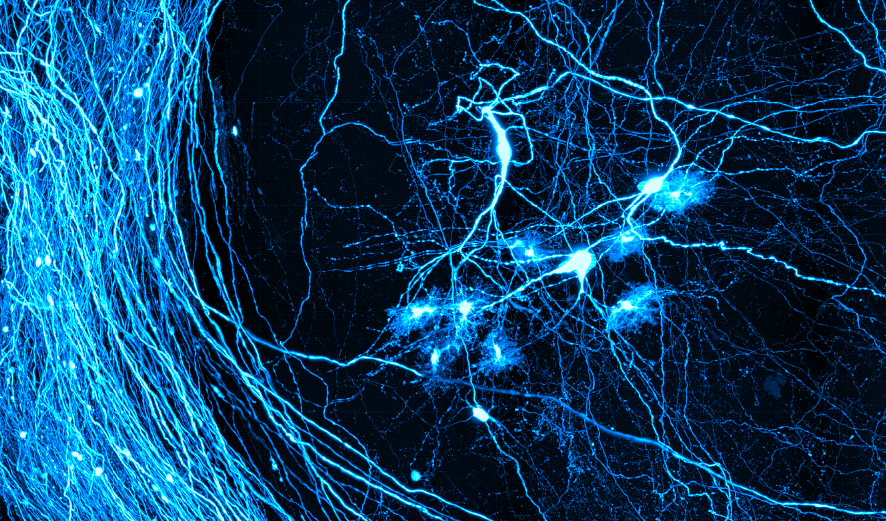
Anderson Chen: The Man Behind the Microscopes


Anderson Chen. Photo by Bernhard Zimmermann.
At the heart of the BU Neurophotonics Center and all of its endeavors lies an important mission: support a range of imaging methods from early stage development to broader adoption and on again to turn-key core facility usage. Meeting this goal involves working with the dozens of photonics faculty at BU who are advancing novel optical imaging technologies—solutions in search of problems, to use the old saying—as well as the many biology faculty who are pursuing research problems that do not yet have a solution. And not only working with the two disciplines, but bridging them and finding ways for the respective investigators to collaborate in as fruitful a way as possible.
In short, furthering the mission is not as easy as it might seem. It takes a particular type of person to understand the gamut of both problems and solutions in play; to envision all the possible ways they might intersect; and finally to support both the developers and users of the optical imaging technologies, to guide the former in adopting their solutions for diverse biological problems and the latter in understanding how to implement those solutions.
Meet Anderson Chen. Trained in experimental physics, Chen is the Senior Imaging Scientist with the Neurophotonics Center and manager of the Micro and Nano Imaging Core facility, which straddles the Neurophotonics Center and the Biomedical Engineering Department at BU. As the Senior Imaging Scientist, he spearheads the building of new microscopes and interacts with biologists about their research. As the manager of the Micro and Nano Imaging Core facility, he interfaces with core users, assists in teaching them how to use the core microscopes, and when needed helps to design their experiments.
“I kind of wear many hats,” he says. Indeed, it is this mix of roles and responsibilities that makes him so integral to the Center and its mission, by facilitating the development of imaging methods from the earliest discussions about the needs the methods will serve to integration into core facilities for simple, turn-key use by a broad swath of biology and other faculty.


Among Chen’s responsibilities at BU is teaching users of the Micro and Nano Imaging Core how best to utilize the microscopes there. Photo by Bernhard Zimmermann.
A True Renaissance Man
Chen’s path to his current position in the Neurophotonics Center was a winding and altogether fascinating one, and in many ways prepared him for the unique set of responsibilities he now has.
After receiving his PhD in experimental physics from Stevens Institute of Technology, in Hoboken, N.J., he began a postdoctoral fellowship at North Carolina State University as a National Research Council scholar. Here, he continued his research with deep sensors, improving the sensors for military applications: developing, for example, a low-cost “hyper-spectral” camera sensor that images the UV through the THz regions on a single, inexpensive imaging chip. (He is quick to point out that the camera bore some resemblance to a device featured in the sci-fi action movie Predator and to the VISOR from Star Trek: The Next Generation.)
The fellowship lasted a year, forcing him to move on, taking a job with the photonics company Newport as a senior electrical optical engineer. The move to industry gave him a new understanding of best practices in bringing developing technologies to market. “I learned a lot about engineering documentation and engineering manufacturing,” he says, “and I gained a new appreciation for time efficiency and the art of design—how to design something that’s easy and cost-effective to build.”
Chen worked with Newport for a year and a half, at the Oriel Instruments Division in Stratford, Connecticut. He might have stayed with the company longer but it was planning to split the division between two existing facilities: one in Irvine, Calif., and the other in Bozeman, Mt. The higher-ups wanted to move him to California but Chen, a street car racer ever since his days in the asphalt jungle of New Jersey, chafed at the idea of moving to a state with such strict emissions standards. Next, they offered him a position in Montana, but “Montana is just too freaking cold and the hail storms would ruin my cars.”
So, in 2014, he moved on again, joining the lab of Na Ji at the Howard Hughes Medical institute Janelia Research Campus in Ashburn, Va. Here he began his journey as a microscopist, developing a simple and robust adaptive optics add-on module for use with two-photon microscopes. “My critical thinking skills sort of took off,” he says. “Dr. Ji really guided me in learning how to formulate processes: how do you establish research questions and how do you answer those questions step by step in microscopy.” His work in the lab was formative, and he might have continued with Dr. Ji when she launched a new lab a couple of years later. But the new lab was in California and, well, the emissions standards.
As the adaptive optics add-on module project at Howard Hughes was winding down, Chen, by happenstance, met David Boas, director of the Neurophotonics Center at BU, at an NIH BRAIN Initiative Conference. Boas was quick to recruit him to join the then-newly formed Center. Thus began the next stage of his journey. Today he looks back fondly on all of the experiences he has had, and all of the mentors and others who have helped to shape him and his career. “Without them,” he says, “I would probably still be hacking away at my little sensor from my PhD. The jobs I’ve had and the people I’ve met along the way have all really helped to expand my horizons.”

Chen’s career path and robust skillset have been shaped by his many, varied interests and experiences. Just one example: His passion for street car racing, which he cultivated in the hidden back streets and industrial stretches of New Jersey, indirectly led him to his current position at BU. Photo by Anderson Chen.
A Better Mousetrap
In addition to training and providing assistance to the many faculty, staff, and students who use the Micro and Nano Imaging Core, Chen is helping to acquire new shared resources for the facility. This past fall, he and other faculty received a “shared instrumentation grant” from the NIH to purchase a new laser scanning confocal microscope. The microscope will replace the one the faculty have been using for the past 12 years, which has now reached the end of its service lifetime, while adding several features previously unavailable to them.
The new instrument will be much faster, capturing images at a video frame rate of almost 30 frames per second on average, as opposed to acquiring an image every two seconds or so with the current instrument. In addition, upgraded detectors in the microscope will also offer improved sensitivity.
That’s not all. Chen’s vision of the facility is to bring cutting-edge equipment and training to the BU campus by partnering with industry leaders. In cooperation with the purchasing department at BU, he negotiated with Olympus, a leading microscope company, to acquire two additional microscopes to complement the new confocal microscope. One of these is a slide scanner that can autonomously and effortlessly image one hundred image histology slides. Previously, this work was generally left to students, who would manually perform the grueling task—which could take many hours to complete and was prone to human errors.
The combination of these new instruments will allow a number of BU faculty to continue their NIH-funded research programs that depend on confocal imaging-based experiments while opening the door to any number of new studies. All of which is good news to Chen, who always appreciates the opportunity to interact with people and learn more about their work. “What makes me especially happy is when I’m able to help them achieve and visualize and finish their projects. The fact that I had a hand in it really speaks to me.”
