The Hardwiring That Makes Us Human
Plato described reason and emotion as two horses pulling the charioteer of the human soul in opposite directions. The divide between these basic human functions persisted throughout philosophy and into psychology and neuroscience, where researchers study the brain’s cognitive and emotive areas independently of one another.
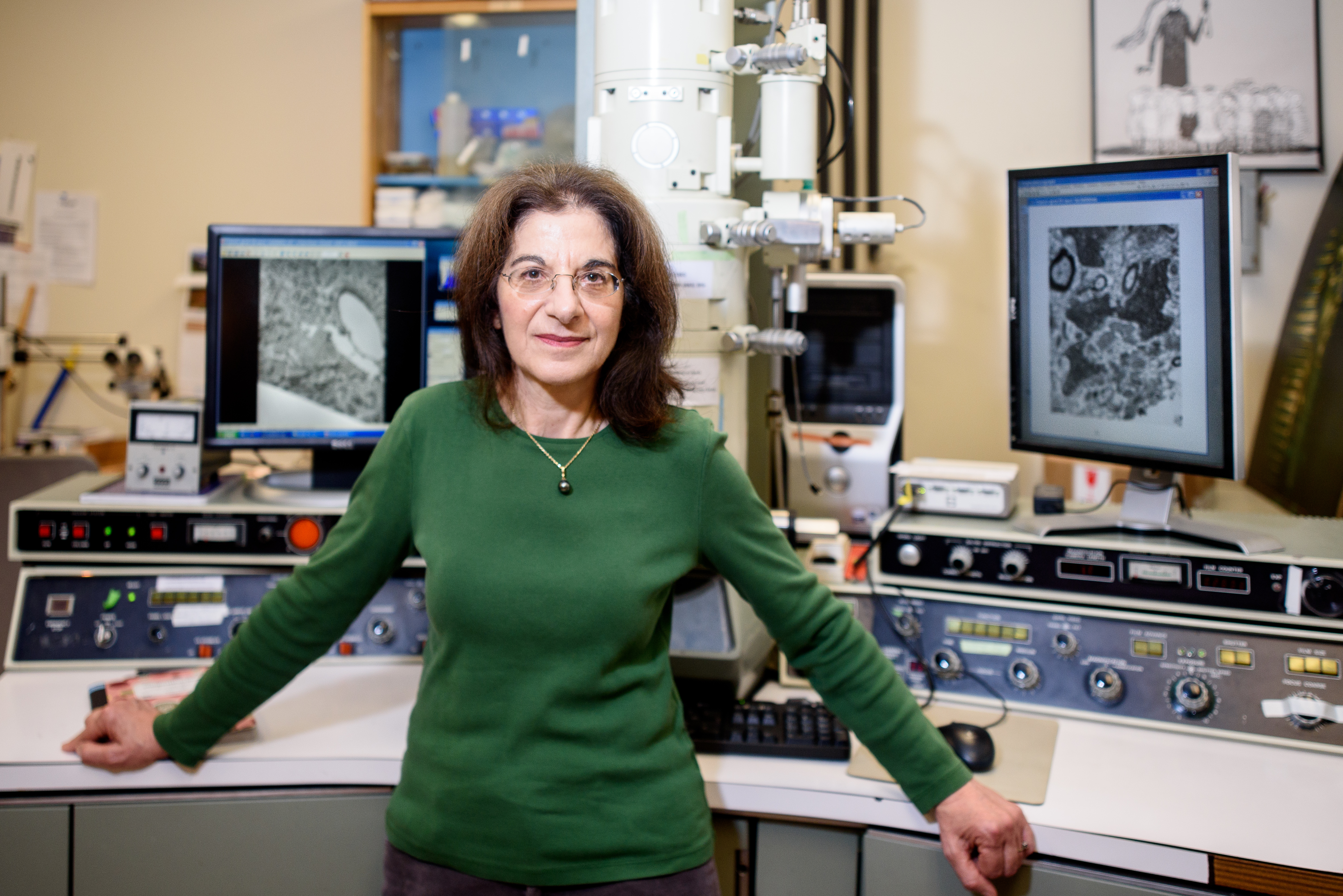
Our cognition, or “what makes us human,” is centered in the prefrontal cortex, the most evolved part of the brain, located just behind our foreheads, says Helen Barbas, a Sargent professor of health sciences. The drives that keep us alive, like hunger and fear, are activated in the hypothalamus and the amygdala, two clusters of neurons deep inside each of the brain’s hemispheres. The amygdala also interprets sensory information; for example, identifying the scent of food or the sound of a predator’s growl.
Researchers tend to consider these areas separately, and “the idea that emotions are not irrational, that they make up a very important part of decision-making, is relatively new” for neuroscience, Barbas says. By mapping the pathways that transmit information throughout the brain, she reveals that the regions for processing emotions and thoughts are inextricably linked—and sundering them “is detrimental to our well-being.”
Until the early 1970s, researchers considered the prefrontal cortex too functionally complex to understand through experimentation. They had found that in a subject under anesthesia, less functionally intricate areas of the brain responded to stimuli; for example, the neurons in the visual area of the brain fired in response to a light shined into the eye. But when the researchers tried to engage the prefrontal cortex, they found it unresponsive and dubbed it the “silent cortex.”
Today, neuroscientists like Barbas know this area is anything but silent. “The prefrontal cortex gets information and does something with it; it’s not just a receiver of information like the sensory areas,” she says. The meaning of the information is important to the prefrontal cortex, and processing it requires consciousness.
“The idea that emotions are not irrational, that they make up a very important part of decision-making, is relatively new” for neuroscience.
—Helen Barbas
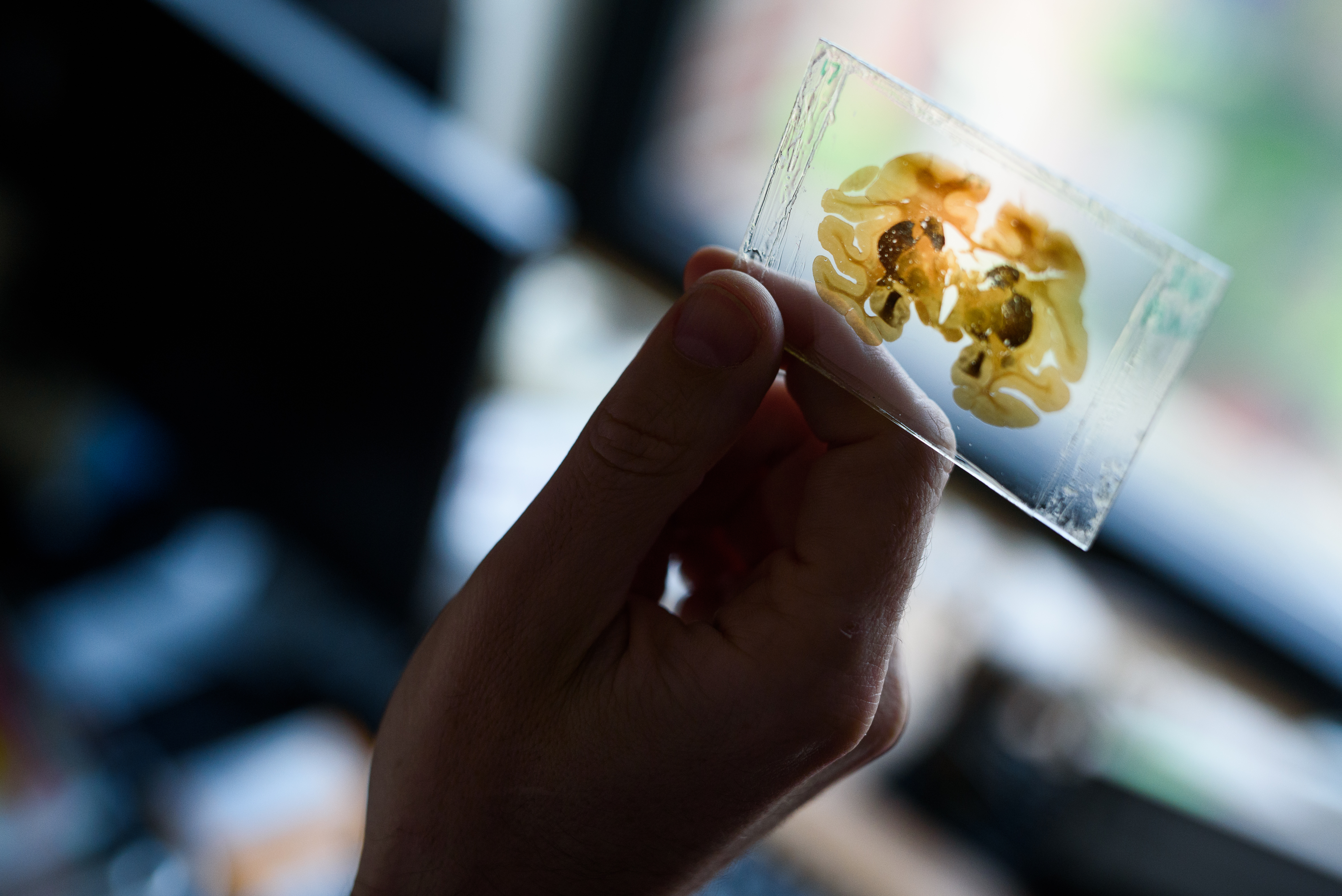
Barbas is one of the first neuroscientists to systematically and quantitatively chart the communication of neurons. She inserts dye into living brain tissue that traces the neurons as they transmit signals throughout the prefrontal cortex and into other areas. By then studying paper-thin slices of the tissue under a microscope, Barbas maps the brain’s communication pathways. Her maps have provided neuroscience with what she calls a bird’s-eye view of this elusive part of the brain.
Let’s say you’re walking along a familiar route when two cars get in a fender bender nearby. You’re likely to jump, then move on. Your response to the accident is determined by the way neurons channel the information about the experience through the brain. That path, Barbas has found, takes an unexpected route.
In a study published in the book The Prefrontal Cortex as an Executive, Emotional, and Social Brain (Springer Japan, 2017), she and co-researcher Miguel Ángel García-Cabezas, a research assistant professor at Sargent, describe how sensory information, like a loud bang, travels deep into the brain to the amygdala, where it is tagged with emotion. Then it heads back out to the prefrontal cortex. “The roundabout way through the amygdala is important to convey information about the significance of an event or a signal,” Barbas says.
When you witness that accident, the amygdala tells your brain that it’s worth noticing, and determines how you’ll react. A person with a typically functioning brain will process the accident as an event of interest, and will go on with their day.
When the pathway between the amygdala and prefrontal cortex is disrupted, however, the brain will likely have a different response to the accident. People with autism, who tend to focus on specific aspects of their environment and are unable to shift their attention to an unexpected event, may not notice the accident. A person with post-traumatic stress disorder (PTSD) may have a panicked response triggered by past trauma.
The ability to shift between emotional states—from startled, back to calm, for example—is dependent on the way neurons transmit information through the amygdala. In a study published in PLOS Computational Biology (2016), Barbas and her colleagues—Assistant Professor Basilis Zikopoulos and Postdoctoral Research Associate Yohan John at Sargent, and Daniel Bullock, a professor of cognitive & neural systems and psychology at BU—modeled pathways between the amygdala, the thalamus, and the prefrontal cortex. These pathways facilitate flexible decision-making, the ability to make quick decisions and change our course of action. If that fender bender blocks your usual way home, you’ll simply find an alternate route.
In another recent article, published in the Journal of Neuroscience (2017) Barbas and her colleagues studied how the pathway from the orbital prefrontal cortex, the part of the prefrontal cortex just behind the eyes, targets different types of inhibitory neurons in the amygdala, specifically those influenced by the regulating molecule dopamine. In a brain with a normal level of dopamine, these inhibitory neurons silence other neurons in the amygdala that are responsible for emotional arousal, keeping us calm. Too much dopamine, and “the system takes over,” Barbas says. That’s why a person with a typically functioning brain who witnesses an accident can return to a state of calm, but a person with PTSD may remain in a state of panic.
“The pattern of connections (between emotion and cognition) can tell us which neurons are more likely to be affected in specific diseases.”
—Helen Barbas
The U-turn the sensory information takes after that fender bender, and the other pathways neurons use to communicate throughout the brain, is determined in part by the brain’s structure, which forms in fetal development, when the areas deep in the brain associated with emotions and cognition “hook up” with the cortex.
Understanding these connections will help researchers and doctors “look at the system in a more mechanistic way and come up with ideas for how to combat neurodegenerative disorders” like PTSD, depression, and schizophrenia, Barbas says. “A disease does not affect all neurons equally. The pattern of connections can tell us which neurons are more likely to be affected in specific diseases.” The next step for Barbas and her colleagues is to study how the brain’s pathways reveal the areas most vulnerable to neurodegenerative diseases, and to determine whether the pathways are responsible for their progression.
Anyone who studies neurological diseases must understand the brain’s circuits to determine where to intervene. For example, many people who have epilepsy are not responsive to drugs and require surgery to remove the affected part of the brain. The surgeon must understand the pathways that will be altered by the procedure so as not to inadvertently disrupt another pathway, impairing speech or causing memory loss, for instance.
That’s why it’s so important for the next generation of researchers to “appreciate how the nervous system is put together and how it may fall apart,” Barbas says. Many undergraduate and graduate students have never seen a brain before they arrive in her lab. “They have seen pictures, but it’s not like the real thing,” she says. “When they work with the brain, they come to appreciate its complexity and how much needs to be done.”
Read more stories from Inside Sargent here.