Chapter 10 – neuroglial cells in general
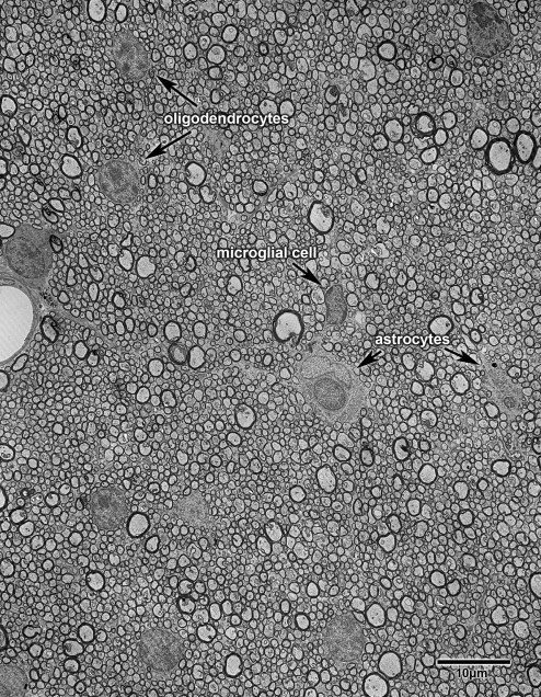
Classically there are three kinds of neuroglial cells, astrocytes, oligodendrocytes and microglial cells (Fig. 10.1).
The population of astrocytes is sometimes subdivided into two categories, protoplasmic and fibrous ones, on the basis of the number, or concentration of filaments in their cytoplasm, as well as the location of the cells. Fibrous astrocytes are those present in white matter, while cytoplasmic ones are most common in gray matter.
Oligodendrocytes have also been classified into categories: (1) interfascicular oligodendrocytes that occur between nerve fibers in white matter; (2) oligodendrocytes in the neuropil of gray matter; and (3) satellite oligodendrocytes that are in close association with neuronal cell bodies. The principal function of the oligodendrocytes is to form myelin, and in the past there has been some dispute about whether or not satellite oligodendrocytes form myelin sheaths. The issue appears to be either unresolved, or of no consequence, the situation being muddied by the fact that using classical methods of staining it was not possible to distinguish between satellite oligodendrocytes and satellite microglial cells.
Satisfactory accounts of the morphology of normal, unreactive microglial cells were not given until the 1970s. One reason for this is that these cells were thought not to exist as a separate entity in the normal brain, but to only appear and to enter the brain under pathological conditions. Another reason is that using the techniques available in early studies these cells were not readily distinguished from oligodendrocytes. In any case it is now agreed that resting microglial cells do exist in the normal brain, making up about 5% of the total population of neuroglia and that with age they become phagocytic.
Myelinating oligodendrocytes in the central nervous system are derived from glial precursors, often referred to as oligodendrocyte precursors, which express two cell surface antigens, proteoglycan NG2 and the platelet-derived growth factor receptor (PDGF aR). In the mature brain, these precursors comprise a fourth type of neuroglial cell population. They make up 4-5% of the neuroglial cell population in the mature brain, and have feature very similar to protoplasmic astrocytes. However, the cytoplasm of these precursors does not contain intermediate astrocytic filaments, and as will be described they have other features that distinguish them from astrocytes.
Figure 10.1
This low power electron micrograph through the anterior commissure of a 6 year old monkey shows the three classical types of neuroglial cells. Oligodendrocytes, which are the most common neuroglial cell type in white matter, have rounded dark nuclei and dark cytoplasm. Astrocytes have pale, somewhat irregularly shaped nuclei and a pale cytoplasm, while microglial cells have dark nuclei that are often kidney shaped.
