Chapter 13 – oligodendrocyte precursor cells
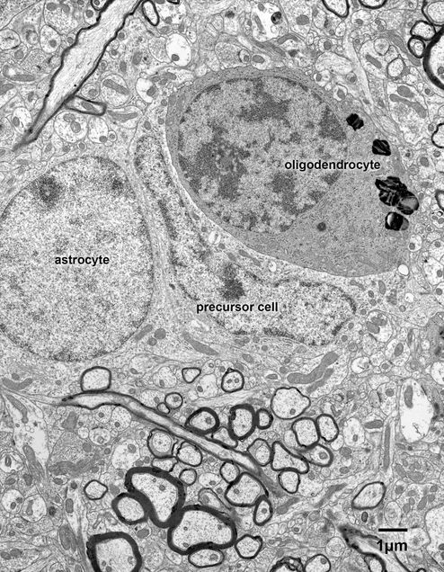
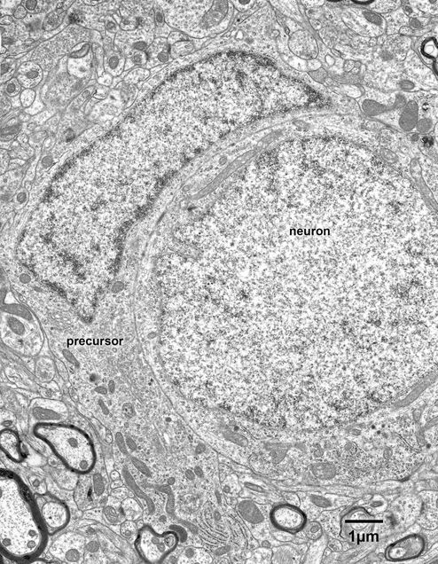
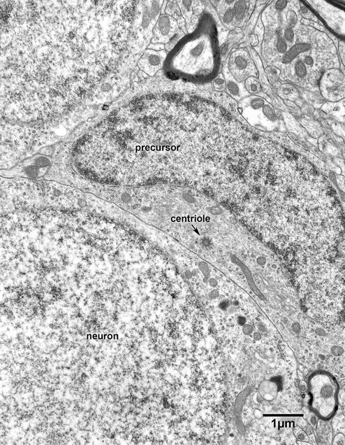
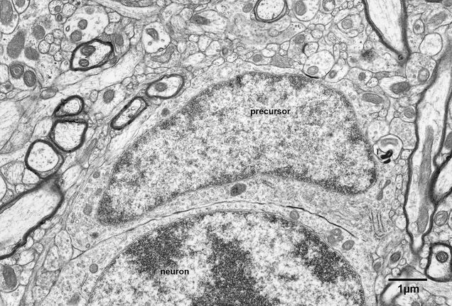
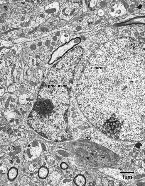
In many respects oligodendroglial precursor cells resemble astrocytes (Fig. 13.1), but their pale nuclei are more irregular is shape than those of astrocytes and they have a more pronounced layer of condensed chromatin beneath the nuclear envelope. Like astrocytes, the cytoplasm of the oligodendrocyte precursors is pale, but the cytoplasm does not contain intermediate filaments (Figs. 13.2- 13.5) and the mitochondria are smaller than those of astrocytes (Fig. 13.1). Although the contours of the cell bodies are irregular, unlike astrocytes the precursors do not send cytoplasmic extensions into the surrounding neuropil and do not mould themselves to the elements in the surrounding neuropil. The only processes arise from the poles of the cell bodies (Figs. 13.2 and 13.2A). In general, the cisternae of rough endoplasmic reticulum in the cytoplasm of precursor cells are short, but there may be stacks of cisternae at the bases of the thick processes extending from the poles of the cell bodies (Fig. 13.2 and 13.2A). It should also be mentioned that although the cytoplasm of these cells may contain a few lysosomes, the cytoplasm never contains inclusions, even in old monkeys (Figs. 13.3 and 13.4).
Figure 13.1
Three neuroglial cells in layer 4 of the primary visual cortex of a 16 year old monkey. The cell on the right is an oligodendrocyte, with the characteristic dark cytoplasm and nucleus. Next to the oligodendrocyte is an oligodendrocyte precursor cell and on the right an astrocyte. Astrocytes and oligodendrocytic precursors have similar features. Both are irregular in shape and their contours fit to the surrounding neuropil, but while the nuclei of astrocytes have rounded profiles, the nuclei of the precursor cells have irregular outlines. Another difference is that the mitochondria of the precursors cells are smaller than those of astrocytes, and the precursor cells never have filaments in their cytoplasm.
Figure 13.1A
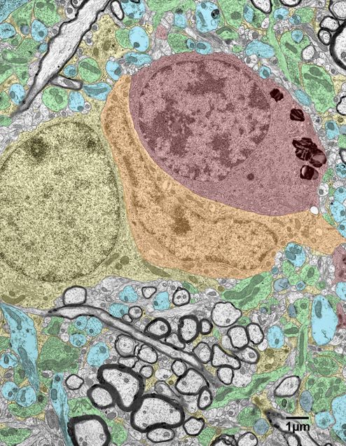
A colored version of Fig.13.1. Oligodendrocytic precursor- orange; oligodendrocyte- red; astrocyte- yellow; neuronal cell bodies and dendrites- blue; dendritic spines- grey; axon terminals- green.
Figure 13.2
An oligodendrocyte precursor cell in the primary visual cortex of a 16 year old monkey. The cell is adjacent to a neuron. Note the irregular shape of the pale nucleus of the precursor cell and the irregular shape of the perikaryon. The perikaryal cytoplasm contains few organelles beyond a few polyribosomes and a few mitochondria, although there are some cisternae of rough endoplasmic reticulum within the process emanating from the cell body.
Figure 13.2A
A copy of the micrograph in Fig. 13.2, in which color has been added. Oligodendrocytic precursor- orange; neuronal cell bodies and dendrites- blue; axon terminals- green.
Figure 13.3
Another example of an oligodendrocyte precursor in the visual cortex of a 25 year old monkey. The precursor is adjacent to the cell body of a neuron. Note the centriole in the cytoplasm of the cell body of the precursor cell.
Figure 13.4
An oligodendrocyte precursor cell adjacent to a neuron in the primary visual cortex of a 35 year old monkey.
Figure 13.5
An oligodendrocyte precursor cell in the primary visual cortex of a 15 year old monkey.
