Chapter 8 – remyelinating axons
It is found that with age there is a significant increase of 20 to 90% in the frequency of profiles of paranodes in both gray and white matter. Since the mean length of paranodes only increases slightly with age, the increase in the frequency of paranodal profiles is attributed to an increase in the number of internodal lengths of myelin with age. This would occur if some of the original lengths of myelin degenerate and are replaced by a series of shorter internodal lengths, as is known to occur in remyelination. And indeed it is generally accepted that the characteristics of remyelination are the presence of myelin sheaths that are inappropriately thin for the size of the enclosed axon and the presence of short internodal lengths of myelin. Both short internodes and inappropriately thin sheaths have been found in old monkeys.
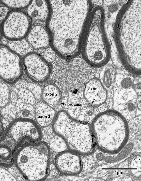
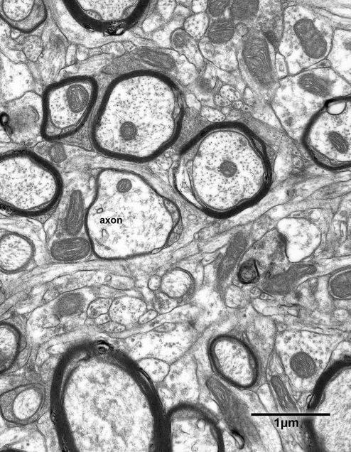
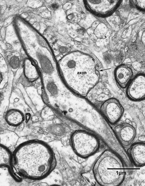
Examples of myelin sheaths that are inappropriately thin for the size of the enclosed axons are shown in Figs. 8.1 – 8.3. In each case the sheath consists of one or two lamellae and the thinness of the sheaths is evident by comparing them with other myelinated fibers in the adjacent neuropil. It is necessary to be careful to ascertain that profiles of paranodes are not mistaken for profiles of thin sheaths, but as pointed out previously, at paranodes the axolemma and the plasma membrane on the inside of the sheath come into close proximity. This is not the case in the three examples of thin sheaths illustrated in Figs. 8.1 – 8.3.
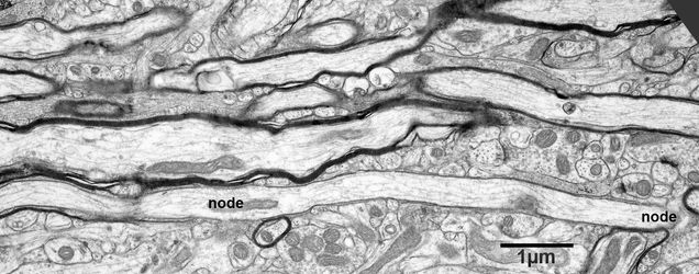
Examples of short internodal lengths of myelin in longitudinal sections of nerve fibers are shown in Figs. 8.4 and 8.5. In both examples the sheaths are very thin and consist of only one or two lamellae.
Figure 8.1
An axon being remyelinated in the primary visual cortex of a 35 year old monkey. The remyelinated axon (axon1) is enclosed by a sheath that appears to consist of a single lamella. The two ends of the process forming the sheath abut at the location indicated by the arrow. Nearby is another axon (axon 2), which is partially surrounded by a process and indicating that it may be in an early stage of remyelination. A third axon (axon 3) is partially covered by a process that is making a junction with its axolemma, suggesting that this axon is sectioned at the level of the paranode/node interface.
Figure 8.2
Neuropil in layer 4 of the primary visual cortex of a 25 year old monkey. One axon (axon) has a sheath that is composed of only one or two lamellae, so that it is inappropriately thin for the size of the enclosed axon. This suggests that the axon is remyelinating.
Figure 8.3
An axon bundle in layer 4 of the primary visual cortex of a 25 year old monkey. One of the axons (axon) is surrounded by a sheath that is only one lamella thick, suggesting that it is in the process of remyelinating. The arrow shows the site where the two ends of the process come together.
Figure 8.4
A bundle of nerve fibers in layer 4 of the primary visual cortex of a 25 year old monkey. One of the nerve fibers is surrounded by a sheath of myelin that is only one lamella thick. The sheath is only three microns long. At each end of the sheath the nodes of Ranvier (nodes) are visible. The thickness of the sheath and the short length of the internode suggest that remyelination is occurring.
Figure 8.5
A bundle of nerve fibers in layer 5 of the primary visual cortex of a 25 year old monkey. One of the nerve fibers is surrounded by a sheath that is only one lamella thick. The internode of myelin is only 5 microns long and the nodes of Ranvier (nodes) are apparent at each end of the internodal length. The short internodal length and the thin sheath suggest that this nerve fiber is undergoing remyelination.


