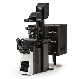
Olympus FV3000 Scanning Confocal Microscope

Olympus FV3000 Inverted Confocal Microscope |

Invitrogen, BPAE Cells, DAPI, Alex488, Mitotracker Red |
Olympus FV3000 is laser scanning confocal microscope to obtain high-resolution, high-contrast images of a sample. It scans samples point by point, resulting in precise 3D fluorescence images.
– 4x Fluorescence detectors, two bi-metal alkali PMT and two high sensitivity GaAsP/GaAs PMT detectors.
– 1x bi-metal Alkali PMT transmission detector for DIC Imaging
– High Efficiency Spectral Detection using Volume Holographic Gratings
– Conventional XY Glavo and High-speed Resonant Galvo-Galvo Scanning
– Motorized XYZ stage for easy transition between macroscopic to microscoping imaging
– 6x solid state diode lasers (405nm, 445nm, 488nm, 514nm, 561nm, 640nm)
– 6x Olympus Objectives
i.) PLAPON, 1.25x, 0.04 NA, 5mm WD
ii.) UPLSAPO10X2, 10x, 0.4NA, 3mm WD
iii.) UPLSAPO20X, 20x, 0.75NA, 0.6mm WD
iv.) LUCPLFLN20X, 20x, 0.45NA, 6.60mm-7.80mm WD
v.) PLAPON60XO, 60x, 1.42 NA, 0.15mm WD (oil dipped)
vi.) UPLSAPO100XS, 100x, 1.35 NA, 0.2mm WD (Si oil dipped)
– Fully enclosed environmental chamber for live cell imaging
– Z Drift Compensation for tracking focal plane shifts during long duration cell imaging trials
– Specialized holders for slides, wellplates, and petri dishes
– CellSense Deconvolution Solution for PSF deconvolution
Acknowledgement Requirements
Users who have gathered data using this microscope must include the following statement in their acknowledgement for any paper, poster and oral presentation.
“Research reported in this publication was supported by the Boston University Micro and Nano Imaging Facility and the Office of the Director, National Institutes of Health of the National Institutes of Health under award Number S10OD024993. The content is solely the responsibility of the authors and does not necessarily represent the official views of the National Institute of Health.”
Other Resources
Quick-Start Guide
Click here to access the Olympus FV3000 Operating Manual Guide. (BU Affiliated members Only)
Click here to access the FV3000 Training Notes REV AD. (BU Affiliated members Only)
Objective Reservation
Click here to request access to the 60x or 100x objectives.
Video Tutorials
Our friends at the UT Dallas has produced some very informative videos on the FV3000. Please visit them at here and like their videos to recognize their work.
Here are their direct links to Youtube.
Olympus Image Format Plugin for ImageJ/Fiji
If you wish to load your saved files into ImageJ, click here to download the ImageJ/Fiji plugin. Once you have downloaded it, please install it like any other plugin for ImageJ and reload ImageJ.
Olympus FV31-SW
Olympus offers free viewing software for the FV3000. Please visit https://www.olympus-lifescience.com/en/support/downloads/ and download the latest version of FV31S-SW Viewer. You can find the FV31-SW software under LASER SCANNING MICROSCOPES.