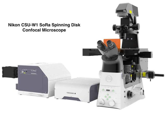
Nikon CSU-W1 SoRA Spinning Disk Confocal Microscope
 |

Invitrogen, BPAE Cells, DAPI, Alex488, Mitotracker Red cells imaged using (Left) 20x, 0.7 NA objective and (Right) 60x, 1.42 NA oil immersion lens with SoRa disk, 4x auxiliary magnification, and Lucy-Richardson deconvolution. |
CSU-W1 SoRa is a NEWLY acquired dual camera spinning disk (inverted) confocal microscope from Nikon, located in the Life Science and Engineering Building (LSEB 222). The confocal imaging platform uses two high-speed Yokogawa spinning disks with series of pinholes to reject out-of-focal plane background signal, providing high quality confocal images at frame rates up to 200 Hz. The CSU-W1 disk has a new pinhole design with optimal pinhole size and spacing, significantly reducing the pinhole crosstalk – an important feature when imaging thicker samples with multiple scattering. The super resolution SoRa disk has additional lenslet array. With high NA objectives and auxiliary magnification, higher quality images with spatial resolution 1.4 the optical limit is possible. Using deconvolution, imaging resolution of CSU-W1 SoRa system can be further increased up to 2x.
Microscope features:
- CSU-W1: Yokogawa spinning disk scanning confocal system for wider field of view and higher image quality
- CSU-W1 SoRa: Spinning disk for super resolution imaging via optical pixel reassignment
- 7 solid state diode lasers (405nm, 445nm, 488nm, 514nm, 561nm, 596nm, and 640nm)
- 2 high sensitivity sCMOS cameras for simultaneous 2-channel imaging
- Motorized XYZ stage for easy transition between macroscopic to microscopic imaging
- High-speed PZT z-translation of z-stack imaging
- 4 Nikon Objectives
i) PLAN APO λD, 4x OFN25, 0.2 NA, 20mm WD
ii) S PLAN FLUOR LWD, 20xC, 0.7 NA, 2.3mm WD
iii) SR PLAN APO IR, 60x, 1.27 NA, 0.18mm WD
iv) PLAN APO λD, 60x, 1.42 NA, 0.15mm WD - Deep learning based algorithm for noise cancellation
- Auxilliary 2.8x or 4x magnification option
- Deconvolution feature for PSF improvement
- Fully enclosed environmental chamber for live cell imaging
- Z Drift Compensation for tracking focal plane shifts during long duration imaging trials
- Specialized holders for slides, wellplates, and petri dishes
Acknowledgement Requirements
Users who have gathered data using this microscope must include the following statement in their acknowledgement for any paper, poster and oral presentation.
“Research reported in this publication was supported by the National Science Foundation Award CBET 2215990. The content is solely the responsibility of the authors and does not necessarily represent the official views of the National Science Foundation.”
Other Resources
- Quick Start Guide / Tutorials (coming soon)
- Nikon offers free viewing software for CSU-W1 SoRa. Click here to download NIS-Element Viewer – a free standalone program to view image files and datasets. The NIS-Elements core packages provide powerful view and image selection modes, including Volume View with 3D Rendering, Tile View for Time, Z, and Multipoint datasets, and Slice View for Z and Time datasets.