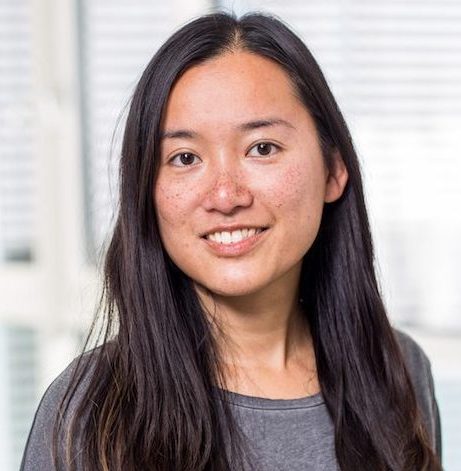
- Title Assistant Professor of Biology; Assistant Professor of Anatomy and Neurobiology
- Education PhD, Albert Ludwig University of Freiburg
- Web Address https://taylab.org
- Email tltay@bu.edu
- Area of Interest neurobiology, neuroimmunology, neurodevelopment, neurodegeneration, traumatic brain injury, brain repair, microglia, glia
- CV
Current Research
How does our brain develop, respond to damage or foreign bodies, and repair itself? The Tay Lab seeks to understand the processes and mechanisms underlying these changes across the lifespan by studying microglia, the principle immune cells in the central nervous system (CNS). Over the past decade, research on microglia have expanded from their immune-sensing functions and disease associations to include their important roles in maintaining brain health and enabling tissue repair. In contrast to neurons and other CNS glial cells derived from the neuroectodermal lineage, microglia primarily originate from a mesodermal source known as the yolk sac erythromyeloid progenitor. Precursors of mature microglia have been observed to infiltrate and establish themselves in the developing brain before the first neuron is formed. Microglia have been described to perform specific activities (e.g., phagocytosis, cytokine release, proliferation) that support the development and maturation of neurons, axons, oligodendrocyte precursor cells and oligodendrocytes, as well as synaptic remodelling and plasticity. Much about the heterogeneity of the microglial populations and their functional diversity remain to be uncovered.
In models of acute neurodegeneration, we have shown that microglia rapidly alter themselves and expand their cell numbers within days in response to the disrupted steady state and to engulf dying neurons or damaged myelin. Clinical recovery and restoration of microglial cell density were observed after four to six weeks. Using a novel reporter mouse model to fluorescently tag microglial cells in different colours, we demonstrated for the first time the removal of excess microglial cells that prevented prolonged inflammation in the brain. While little is known about the underlying molecular signals controlling the restoration of normal microglial cell distribution and homeostatic phenotypes, our single-cell transcriptomic approach revealed a specific gene signature associated with the onset of recovery that will be investigated in greater detail. Collectively, the evidence is clear that microglia are critical drivers of brain tissue repair. We want to find out how they do their job, and in the case of chronic neurodegeneration, why they fail to do their job.
We will apply a wide array of approaches, including the use of different biological models (e.g., cell cultures, tissues, organisms), high-resolution microscopy, next-generation sequencing, multi-omic technologies, and machine learning-based analyses. We form extensive interdisciplinary collaborations to broaden our range of research tools, including the development of neural microelectrodes with microsystems engineers, and to strengthen the translational aspects of our research. Through understanding the heterogeneity, fundamental biological mechanisms, functions, and intercellular interactions of microglia, we hope to provide insights that alleviate neuropsychiatric disorders and neurodegenerative diseases, including addiction, anxiety, major depression, stress, dementia, Alzheimer’s disease, and traumatic brain injury.
Selected Publications
- Paolicelli, R. C., Sierra, A., Stevens, B., Tremblay, M-È, … Wyss-Coray, T. (2022). Microglia states and nomenclature: A field at its crossroads. Neuron, 110 (21): 3458-3483.
- Mehl LC, Manjally AV, Bouadi O, Gibson EM, and Tay TL (2022) Microglia in Brain Development and Regeneration. Development, 149 (8): dev200425.
- Costa Jordão MJ, Brendecke SM, Sankowski R, Sagar, Locatelli G, Tai Y-H, Tay TL, Schramm E, Armbruster S, Hagemeyer N, Mai D, Çiçek Ö, Falk T, Kerschensteiner M, Grün D, Prinz M (2019) Single-cell profiling of the myeloid compartment identifies new cell populations with distinct fates during neuroinflammation. Science, 363: eaat7554.
- Falk T, Mai D, Bensch R, Çiçek Ö, Abdulkadir A, Marrakchi Y, Böhm A, Deubner J, Jäckel Z, Seiwald K, Dovzhenko A, Tietz O, Dal Bosco C, Walsh S, Saltukoglu D, Tay TL, Prinz M, Palme K, Simons M, Diester I, Brox T, Ronneberger O (2019) U-Net: deep learning for cell counting, detection, and morphometry. Nat Methods, 16: 67-70.
- Shemer A, Grozovski J, Tay TL, Tao J, Süß P, Volaski A, Gross M, Kim J-S, David E, Chappell-Maor L, Thielecke L, Glass CK, Cornils K, Prinz M, Jung S (2018) Engrafted parenchymal brain macrophages differ from host microglia in transcriptome, epigenome and responsiveness to challenge. Nat Commun, 9: 5206. (co-first author)
- Tay TL, Sagar, Dautzenberg J, Grün D, Prinz M (2018) Unique microglia recovery population revealed by single-cell RNAseq following neurodegeneration. Acta Neuropathol Commun, 6: 87. (co-corresponding and co-first author)
- Tay TL, Mai D, Dautzenberg J, Fernandez-Klett F, Lin G, Sagar, Datta M, Drougard A, Stempfl T, Ardura-Fabregat A, Staszewski O, Margineanu A, Sporbert A, Steinmetz L, Pospisilik JA, Jung S, Priller J, Grün D, Ronneberger O, Prinz M (2017) A new fate mapping system reveals context-dependent random or clonal expansion of microglia. Nat Neurosci, 20(6): 793-803. (co-corresponding author)
- Tay TL, Ronneberger O, Ryu S, Nitschke R, Driever W (2011). Comprehensive catecholaminergic projectome analysis reveals single-neuron integration of zebrafish ascending and descending dopaminergic systems. Nat Commun, 2: 171.