Miniature Computational Imaging System Advances Brain Imaging Technology
The ability to study and learn about the brain hinges on what technology is available. CISE faculty affiliate, Lei Tian (ECE, BME) is leading the development of a novel miniature computational imaging system to advance brain imaging technology.
Tian (ECE, BME), was awarded a $2 million grant from the National Institutes of Health to advance the development of the Computational Miniature Mesoscope (CM2). The CM2 is a “wearable” miniaturized neural imaging device that Tian has been developing in collaboration with Professors David A. Boas (BME, ECE) and Ian G. Davison (Biology) since the concept was first funded through a Dean’s Catalyst award in 2018, internal support from BU Neurophotonics Center, and later prototyped through a subsequent NIH BRAIN initiative R21 grant.
“With this R01 grant, we want to take CM2 from a proof-of-concept prototype to a more mature technology so that it can be broadly impactful and adopted in the neuroscience community,” says Tian.
One issue with the current technology is that the imaging devices are still too large and heavy for small animals like mice to wear while moving freely during the in-vivo neural recording. An important component of CM2 is its wearability, which will allow scientists to record neuronal activities on freely moving mice. For this to be successful, the device needs to be extremely light and can weigh only < 4 grams.
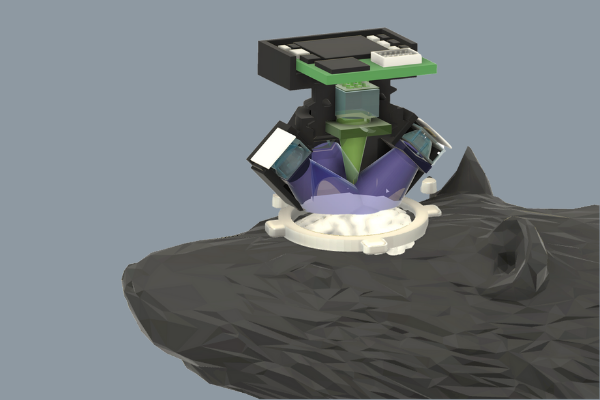
Across the cortex of the mouse, there are tens of thousands of neurons firing in distinctive patterns depending on the brain’s activity. The project aims to capture a complete picture of brain activity across the entire mouse cortex while maintaining single-neuron resolution. The technology explores miniature optics and advanced computational algorithms to achieve novel capabilities beyond conventional technologies. To enable imaging across multiple spatial scales, the device uses an array of microlenses, inspired by insect eyes, to provide a full-cortex, high-resolution picture of the cortex, and enables 3D imaging to accommodate for the cortical curvature. “Typically, these are competing factors,” says Tian. “Images either have a high resolution or a wide field of view. We’re using the computational imaging approach to overcome these tradeoffs.”

The CM2 will capture calcium dynamics expressed in fluorescence intensities emitted from the brain of transgenic mice. A critical part of Tian’s project lies in using highly efficient LED light to excite the fluorescence. The CM2 presents a novel design, using an LED array and 3D-printed freeform optics to excite fluorescence with high light efficiency throughout the entire cortical surface.
The team working on CM2 has a multidisciplinary background which has enabled them to develop the innovative technology demonstrated in CM2. The team features Principal Investigator Tian, who has expertise in computational microscopy and deep learning for biomedical optics, co-Investigator Boas, whose background is in biomedical optics and neurophotonics, and co-Investigator Davison, an expert in wearable microscopes. “There needs to be a bridge between technology and application,” Tian explained. “Boas and Davison have the unique expertise to help translate the technology from my optics lab to a device that is attractive to the broad neuroscience community.”
Read more about the project here.