FAQ
COVID-19 Information
- Our return-to-work plan.
- Symptom Screening
- Checklist for day of cleaning and safety protocols.
- IRB information.
- BU’s central repository for returning to campus during this time.
- Environmental Health and Safety information – including cleaning policies and return-to-work information.
Getting Started
- How to sign up for the mailing list (cncmri-list).
- How do I get access to the center?
- Badge system for center access.
- MRI safety training information.
- How do I get access to the testing rooms?
- How do I get access to the scheduling website?
- Where do I go to analyze my data – number crunching!
General Information
- MRI Screening Form
- What should I cite and acknowledge for work conducted at the CNC?
- How do I write the scanning parameters for my paper – an example methods section.
- Past workshop recordings, including Intro to AFNI, SPM, Matlab, MR physics, Eyelink, Freesurfer, UNIX and more!
SCC Cluster Questions
- How do I get an account on the SCC?
- How do I manage my account on the SCC – add more space/users?
- How do I get on the SCC?
- How do I access OnDemand to get on the SCC?
- General help for OnDemand
- OnDemand FAQ including:
- When I start my app in OnDemand, I can not see the menu for my application and I can’t see the title bar in order to move the window. What should I do?
- How do I Copy & Paste from within OnDemand?
- How do I change the default keyboard shortcuts (e.g. copy/paste) in the SCC OnDemand Desktop?
- How do I get my dicoms from XNAT to the SCC?
- How do I select software on the cluster?
- Any tips for using R, Python, Matlab, C, Fortran on the SCC?
- How do I submit a job on the cluster- qsub?
Scanner Related Questions
- How do I get the trigger and button box responses?
- How to put condition markers into the Biopac trace.
- How to use the video grabber to see if the eye is open.
How to sign up for the mailing list
All mri users should be on the mailing list. This is where we will announce issues with the equipment, training opportunities, and procedural updates. It is also a place where people can share information they think might be useful to the group, or send out a request for participants. Though please try to keep the email traffic to as little as possible. This is a majordomo list that does not have a web interface. To sign up for it you must send the list an email. Follow the rules below:
1. Send an email to majordomo@bu.edu
2. The body of the message has to include:
subscribe cncmri-list@bu.edu your_email_address@blah.blah
3. It has to be sent in plain text. This is usually not how most people send emails these days. For instructions on changing to plain text see here.
4. It cannot contain a signature, or anything else in the body of the email.
5. It cannot contain a subject line.
How do I get access to the center?
Access to the Cognitive Neuroimaging Center is restricted by card readers posted at multiple entrances and positions within the facility and is based on a badge system. Individuals affiliated with the center, who have completed appropriate human subjects training, and badge requirements will be granted access.
To request swipe card access to the CNC (using your standard issue BU ID card), please fill out the form here, AND send a notification email to the CNC staff, Shruthi Chakrapani and Stephanie McMains (schakra@bu.edu and mcmains@bu.edu) When filling out the form, choose Cognitive Neuroimaging Center from the list of centers / PIs, and fill out the remaining information. Requests will be processed as quickly as possible, but may take up to 3 business days to complete.
If you will be using the scanner, please make sure the CNC is listed as a scanning site on your IRB.
Badge system for center access.
Users are required to obtain and display a badge with photographic identification when using the Cognitive Neuroimaging Center facilities. Several different badges may be obtained, depending on access requirements, and contingent on level of training. Access levels to the center are described briefly in the table below.
To request a badge, please fill out the online form here.
| BADGE COLOR | ACCESS LEVELS | ELIGIBILITY CRITERIA | HOW TO |
| RED |
BU ID access to the center entrance door Access to corridor testing rooms NO ACCESS TO SCANNER SUITE |
Completed human subjects training Approval by CNC staff |
Fill out the form here to request center access |
| YELLOW |
All red badge privileges, and: Access to scanner suite while escorted by a green badge user or CNC staff |
All red badge criteria, and: Must complete MR safety training course Must pass CNC online safety quiz (80%+) |
Express interest in a future safety training course here A link to study materials and an online quiz will be provided after training |
| GREEN |
All yellow badge privileges, and: BU ID access to the scanner suite Can lead scan sessions during business hours (M-F 9:00-5:30) while CNC staff is present at the center |
All yellow badge criteria, and: Completion of 10 independent scanning sessions under the supervision of a green or silver badge user or CNC MRI technologist Schedule and complete a scanner training session with CNC MRI technologist Pass oral quiz with CNC MRI technologist |
Keep a log of your training sessions as a yellow badge user Email Shruthi (schakra@bu.edu) to schedule a training session or quiz |
| SILVER |
All green badge privileges, and: Can lead scan sessions off hours (weekdays 5:30-9:00 pm and weekends 9:00-5:00pm) without CNC staff present at the center |
All green badge criteria, and: Should be an experienced (2+ years) green badge user Should have completed a minimum of 10 independent scan sessions at CNC without supervision Pass oral quiz with CNC MRI technologist |
Silver badge eligibility will be determined on a case-by-case basis. Those interested in obtaining this status can contact CNC staff to discuss |
MRI safety training information.
Currently, MRI Safety Training sessions are scheduled as needed. To express interest in attending a session, please fill out the online form here.
All users who wish to access the Cognitive Neuroimaging Center MRI Suite must have completed relevant human subjects training and also must complete an in-person MRI safety training course. The CNC staff offers a semi-regular 2-hour safety training session, during which users will be presented with the fundamental issues related to MR safety and procedures to follow in our center. The training also includes a brief walk-through of the scanner suite. After the training session, users will receive a link to the materials presented and must complete and pass (80% correct or greater) an online quiz.
How do I get access to the testing rooms?
Please email Shruthi and include information about your PI and the rooms you need access to.
How do I get access to the scheduling website?
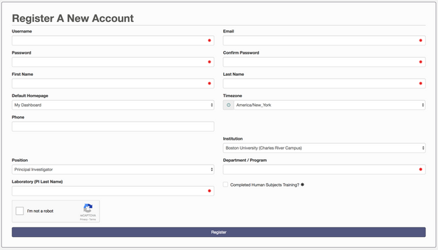
First you need to go to the website and click on Register.
Next, email Shruthi (schakra@bu.edu) with what calendar you need access to (testing room, fNIRS, MRI).
Upon registration, accounts will be verified by CNC staff and added to the CNC User groups requested, which will enable scheduling of resources.

You are required to check the box next to Completed Human Subjects Training. Please make sure you have completed your training before requesting access.
General intro to the scheduling system.
The easiest way to interact with the system is from the Resource Calendar view. From the menu across the top choose Schedules -> Resource Calendar.
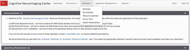
There will then be a dropdown menu above the calendar. You can use this to select the calendar you are interested. The three main categories are (1) MRI Suite (Business Hours), (2) MRI Suite (Off Hours), and (3) Testing Rooms (24/7 availability). Not all users will have access to the MRI calendars, and not all MRI users will have access to the Off Hours calendar. Please contact Shruthi for access to additional calendars.
Importantly, choose the week view to give a better idea of how long each booking is. The month view can be deceiving.
How to book a testing room.
Make sure you are viewing the correct calendar, and then Click on the day you want. You will get a choice to create a reservation, view the day, or cancel – this just closes the popup menu.
Choose create a reservation and verify the date and set the time. You can make a reservation for a single instance, or to repeat at different intervals.
The Project Name field will be what shows up in the calendar. This should be something short indicating the lab and possibly a project name.
Testing room reservations do not require approval and can be made at any time.
How to book MRI scan time.
Currently all MRI suite reservations require approval from CNC staff, and must be made at least 24 hours in advance. These reservations will thus be in a pending state until approved by staff.
Make sure you are viewing the correct calendar, and then Click on the day you want. You will get a choice to create a reservation, view the day, or cancel – this just closes the popup menu. Choose create a reservation, it will bring up the form below.
How to fill out the reservation form
Verify the date and start/end times.
Title: Often the XNAT project, or lab name, up to you.
Description: Can be left empty for most. If you are doing testing, or anything that is different than normal, that may affect whether you need assistance, please list it here.
Project Name: This should be your XNAT project, for outside users it can be your study name. This is what shows up in the calendar.
IRB Protocol and Funding Source are required. If you have questions, email Shruthi.
Will 2+ MRI-trained personnel be present? This has a drop down menu, shown below.

Using the MRI suite currently requires two MRI trained personnel to be present in the scanner suite throughout the session, as well as the presence of a member of the CNC staff or a PI with MRI training inside the center (see silver badge exception).
Yes(including 1+ green badges)
If you have at least one green badge, and a second person with at least a yellow badge.
Yes(1 silver & 1 green/off hours ok)
If you have a silver badge and a green badge, you are not required to have the CNC staff in the center. This allows you to scan off hours, or at other times when no staff is present. Please choose if you have the required people even if you don’t need it (i.e. scanning during regular hours).
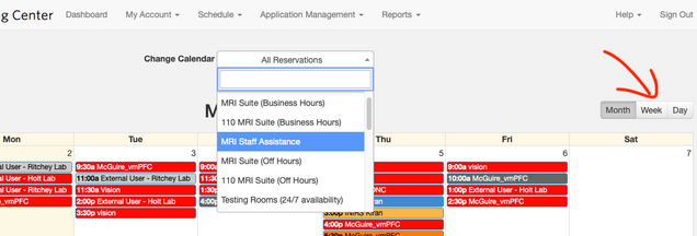
Yes (2 yellow badge/requested MRI Staff Assist
If one of the two trained personnel is not a green badge (i.e. you have two yellow badges), then you require assistance from the CNC staff for your session. You should book both the MRI suite and the MRI Staff Assistance resources for your session. Click on Change, next to resources, it will bring up a window, select both 110 MRI Suite and MRI Staff Assistance.
![]()
No (No participant, just testing)
Depending on what testing you are doing, you might need staff assistance. If you are just testing equipment, like button boxes, you generally don’t. If you have to put a phantom in and run the scanner, like if you are testing the trigger, then you need help if you aren’t a green badge. Please utilize the Description of Reservation field.
Where do I go to analyze my data – number crunching!
Boston University supports the SCC, a shared Linux cluster for high-performance computing. See their detailed description: https://www.bu.edu/tech/support/research/computing-resources/scc/.
Please see our additional help topics on the FAQ: https://www.bu.edu/neuroimaging/information-for-investigators/faq/scc-cluster-questions/.
What should I cite and acknowledge for work conducted at the CNC?
If you collected data on the MRI scanner, you should acknowledge the NSF grant. If you used ONR compute resources, then you should acknowledge the ONR grant. If you used SMS/multiband sequences, you should acknowledge the CMRR and include several citations related to the SMS/Multiband techniques. If you use iPAT then there is also a citation to add. See an example acknowledgement below:
This research was carried out at the Boston University Cognitive Neuroimaging Center. This work involved the use of instrumentation supported by the NSF Major Research Instrumentation grant BCS-1625552. We acknowledge the University of Minnesota Center for Magnetic Resonance Research for use of the multiband-EPI pulse sequences. Data was analyzed on a high-performance computing cluster supported by the ONR grant N00014-17-1-2304.
These are the citation you should include. To see an example of how to use them, see the example methods section below.
van der Kouwe A.J.W., Benner T., Salat D.H., Fischl B. (2008). Brain morphometry with multiecho MPRAGE. NeuroImage, 40, 559–569.
Moeller S., Yacoub E., Olman C.A., Auerbach E., Strupp J., Harel N., Uğurbil K. (2010). Multiband multislice GE-EPI at 7 Tesla with 16-fold acceleration using Partial Parallel Imaging with application to high spatial and temporal whole-brain FMRI, Magn. Reson. Med., 63, 1144–1153.
Feinberg D.A., Moeller S., Smith S.M., Auerbach E.J., Ramanna S., Glasser M.F., Miller K.L., Ugurbil K., Yacoub E. (2010). Multiplexed Echo Planar Imaging for Sub-Second Whole Brain FMRI and Fast Diffusion Imaging, PLoS One, 5:e15710.
Setsompop K., Gagoski B.A., Polimeni J.R., Witzel T., Wedeen V.J., Wald L.L. (2012). Blipped-controlled aliasing in parallel imaging for simultaneous multislice echo planar imaging with reduced g-factor penalty, Magn. Reson. Med., 67,1210-1224.
Xu, J., Moeller S., Auerbach E.J., Strupp J., Smith S.M., Feinberg D.A., Yacoub E., Ugurbil K (2013). Evaluation of slice accelerations using multiband echo planar imaging at 3 T. Neuroimage, 83, 991-1001.
Cauley S., Polimeni J., Bhat H., Wald L., Setsompop K. (2014). Interslice leakage artifact reduction technique for simultaneous multislice acquisitions. Magn Reson Med, 72(1):93-102.
For iPAT:
Griswold, M., Jakob, p., Heidemann, R., Nittka, M., Jellus, V., Wang, J., Kiefer, B., Haase, A. (2002). Generalized autocalibrating partially parallel acquisitions (GRAPPA). Magn Reson Med., 47(6):1202-10.
How do I write the scanning parameters for my paper – an example methods section.
You should update the individual parameters to match what you used. If you need help getting the correct numbers, feel free to email Shruthi (schakra@bu.edu).
Data were collected on a 3T Siemens Prisma MRI scanner (Siemens Healthcare, Erlangen, Germany) using the vendor’s 64-channel head coil. The Siemens Auto-Align tool was used to ensure reproducible placement of image fields of view. Anatomical images were collected with a T1-weighted magnetization-prepared rapid gradient multi-echo sequence (multi-echo MPRAGE [1], 176 sagittal slices, TR = 2530 ms, TEs = 1.67, 3.5, 5.33, and 7.16 ms, TI = 1100 ms, flip angle = 7°, 1 mm3 voxels, FOV = 230 mm, GRAPPA (iPAT) [2] acceleration = 4). All Blood-oxygen-level-dependent (BOLD) data were collected via a T2*-weighted echo-planar imaging (EPI) pulse sequence that employed multiband RF pulses and Simultaneous Multi-Slice (SMS) acquisition [3-7]. For the task runs, the EPI parameters were: 75 interleaved axial-oblique slices (25 degrees toward coronal from ACPC alignment), TR = 2000 ms, TE = 28 ms, flip angle = 80°, 2.0 mm isotropic voxels, FOV = 208 mm, SMS factor = 3, GRAPPA acceleration = 2). The SMS-EPI acquisition used the CMRR-MB pulse sequence from the University of Minnesota.
Notes
- FOV: This is usually the FOV read. It is sometimes written as 230 x 230 mm. When it is written this way, it is usually the FOV read x FOV write. And you only need this if they differ, and it is not mm squared.
Citations
Please ensure all the references are cited regardless of how you write the parameters.
- van der Kouwe A.J.W., Benner T., Salat D.H., Fischl B. (2008). Brain morphometry with multiecho MPRAGE. NeuroImage, 40, 559–569.
- Griswold, M., Jakob, p., Heidemann, R., Nittka, M., Jellus, V., Wang, J., Kiefer, B., Haase, A. (2002). Generalized autocalibrating partially parallel acquisitions (GRAPPA). Magn Reson Med., 47(6):1202-10.
- Moeller S., Yacoub E., Olman C.A., Auerbach E., Strupp J., Harel N., Uğurbil K. (2010). Multiband multislice GE-EPI at 7 Tesla with 16-fold acceleration using Partial Parallel Imaging with application to high spatial and temporal whole-brain FMRI, Magn. Reson. Med., 63, 1144–1153.
- Feinberg D.A., Moeller S., Smith S.M., Auerbach E.J., Ramanna S., Glasser M.F., Miller K.L., Ugurbil K., Yacoub E. (2010). Multiplexed Echo Planar Imaging for Sub-Second Whole Brain FMRI and Fast Diffusion Imaging, PLoS One, 5:e15710.
- Setsompop K., Gagoski B.A., Polimeni J.R., Witzel T., Wedeen V.J., Wald L.L. (2012). Blipped-controlled aliasing in parallel imaging for simultaneous multislice echo planar imaging with reduced g-factor penalty, Magn. Reson. Med., 67,1210-1224.
- Xu, J., Moeller S., Auerbach E.J., Strupp J., Smith S.M., Feinberg D.A., Yacoub E., Ugurbil K (2013). Evaluation of slice accelerations using multiband echo planar imaging at 3 T. Neuroimage, 83, 991-1001.
- Cauley S., Polimeni J., Bhat H., Wald L., Setsompop K. (2014). Interslice leakage artifact reduction technique for simultaneous multislice acquisitions. Magn Reson Med, 72(1):93-102.