Ed Boyden

Ed Boyden is Y. Eva Tan Professor in Neurotechnology at MIT, professor of Biological Engineering and Brain and Cognitive Sciences at MIT’s Media Lab and McGovern Institute for Brain Research, and was recently selected to be an Investigator of the Howard Hughes Medical Institute (2018). He leads the Synthetic Neurobiology Group, which develops tools for analyzing and repairing complex biological systems such as the brain, and applies them systematically to reveal ground truth principles of biological function as well as to repair these systems. These technologies include expansion microscopy, which enables complex biological systems to be imaged with nanoscale precision; optogenetic tools, which enable the activation and silencing of neural activity with light; robotic methods for directed evolution that are yielding new synthetic biology reagents for dynamic imaging of physiological signals, such as neural voltage; novel methods of noninvasive focal brain stimulation; and new methods of nanofabrication using shrinking of patterned materials to create nanostructures with ordinary lab equipment. He co-directs the MIT Center for Neurobiological Engineering, which aims to develop new tools to accelerate neuroscience progress.
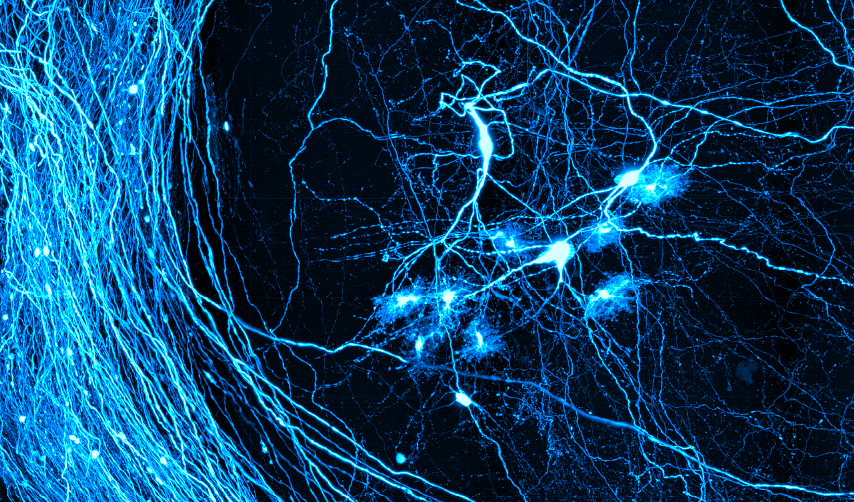
Dr. Boyden will be leading a discussion on Optical Tools for Analyzing and Repairing Complex Biological Systems. Understanding and repairing complex biological systems, such as the brain, requires new technologies that enable such systems to be observed and controlled with great precision, across extended spatial and temporal scales. He and his team are discovering new molecular principles that are leading to such technologies. For example, they recently discovered that it was possible to physically magnify biological specimens manyfold, in an even way, by embedding them in dense swellable polymers, mechanically homogenizing the specimens, and then adding water to isotropically swell the specimens. In this method, which they call expansion microscopy (ExM), they enable scalable, inexpensive diffraction-limited microscopes to do large-volume nanoscopy, in a multiplexed fashion – important, for example, for brain mapping. As another example, they discovered that microbial opsins, genetically expressed in neurons, could enable their electrical activities to be precisely driven or silenced in response to millisecond timescale pulses of light. These tools, called optogenetic tools, are enabling causal assessment of the contribution of defined neurons to behaviors and pathologies in a wide variety of basic science settings. Finally, they have developed new methods of directed evolution, and discovered mutant forms of optogenetic tools that enable precision fluorescent imaging of the high-speed voltage of neurons in the living brain. They share all these tools freely, and aim to integrate the use of these tools so as to lead to comprehensive understandings of neural circuits.