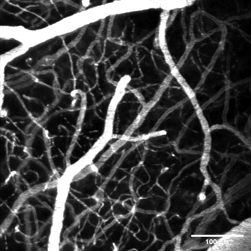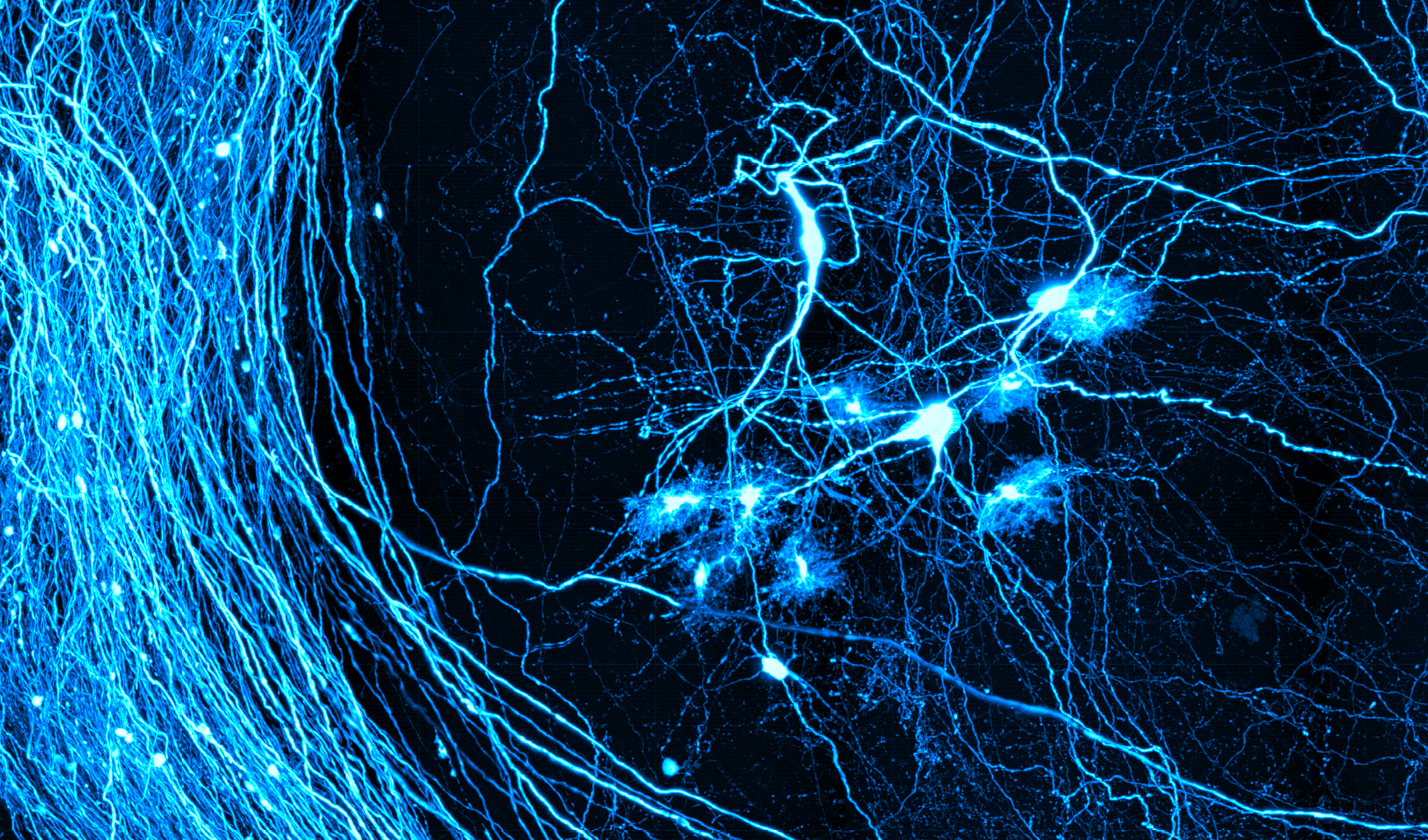
Multiphoton Microscopy
Nonlinear excitation of fluorescence allows for excellent optical sectioning, enabling high resolution imaging deep into highly scattering tissue. Two-photon microscopy is one of the most widely used tools for neuroimaging, letting researchers image dynamics such as neural activity and blood flow deep into the brain.
The Neurophotonics Center has a commercial system (Bruker Investigator) available for use. The system has a tunable excitation source and multiple filter sets to suite a variety of applications. It provides not only standard two photon-imaging but also includes a resonant scanning module for video rate imaging as well as a TCSPC module for fluorescent lifetime imaging (FLIM).

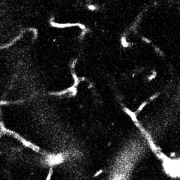
Bessel Beam
Our custom two photon system includes an additional module creating an elongated Bessel-like focus. By extending the axial focus, a large volume can be imaged at high lateral resolution with a single 2D raster scan. This allows us to image volumes up to 713x713x120μm at the same frame rate of a standard two photon microscope. We are also in the process of integrating and OCT system for multimodal imaging studies.