MicroCT Imaging and 3-D X-Ray Microscopy
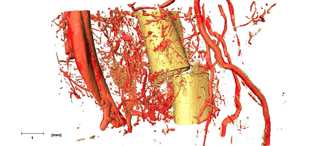


Micro-computed tomography (μCT) is a three-dimensional, X-ray-based imaging modality that can image tissues, organs and whole organisms as well as nonorganic structures with a spatial resolution as high as 6-10 μm. μCT systems provide rapid, quantitative, high-resolution and three-dimensional assessment of both microstructure and density. μCT is nondestructive and can scan ex-vivo samples as large as 75 cubic centimeters in several hours.
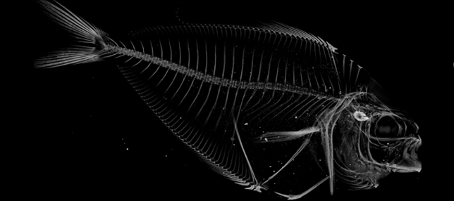
X-ray microscopy (XRM) is a nearly identical imaging modality that combines X-ray imaging with microscope technology to enhance the spatial resolution to 0.7 μm. This unique set-up allows for ex-vivo and nonorganic samples of almost any size or shape to be imaged.
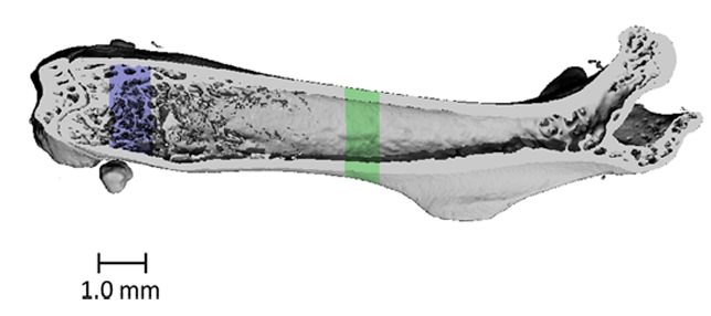
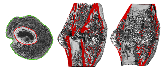
The Micro-Computed Tomography Imaging Core Facility at BU has one μCT scanner and one X-ray microscope. The μCT scanner is a Scanco μCT40 system which is density calibrated on a weekly basis to keep our scans accurate. The XRM is the Zeiss Xradia Versa 520. We have multiple software options that allow for the identification of sub-regions for image segmentation, registration, and quantitative analysis. Analyses can be performed on three-dimensional regions of interest to quantify porosity, thickness, and other features of the microstructure. Additionally, two-dimensional regions of interest can be analyzed to quantify bone area, bone area fraction, cross-section area, and moment of inertia. We have the ability to render and export images in a variety of file formats.
The systems are available for use regardless of affiliation with Boston University. The cost per scan roughly scales with the amount of image data that are generated as well technician consultation, and optional analyses performed. Prospective users should contact the lab via the imaging contact form.