Past Beckman Scholars
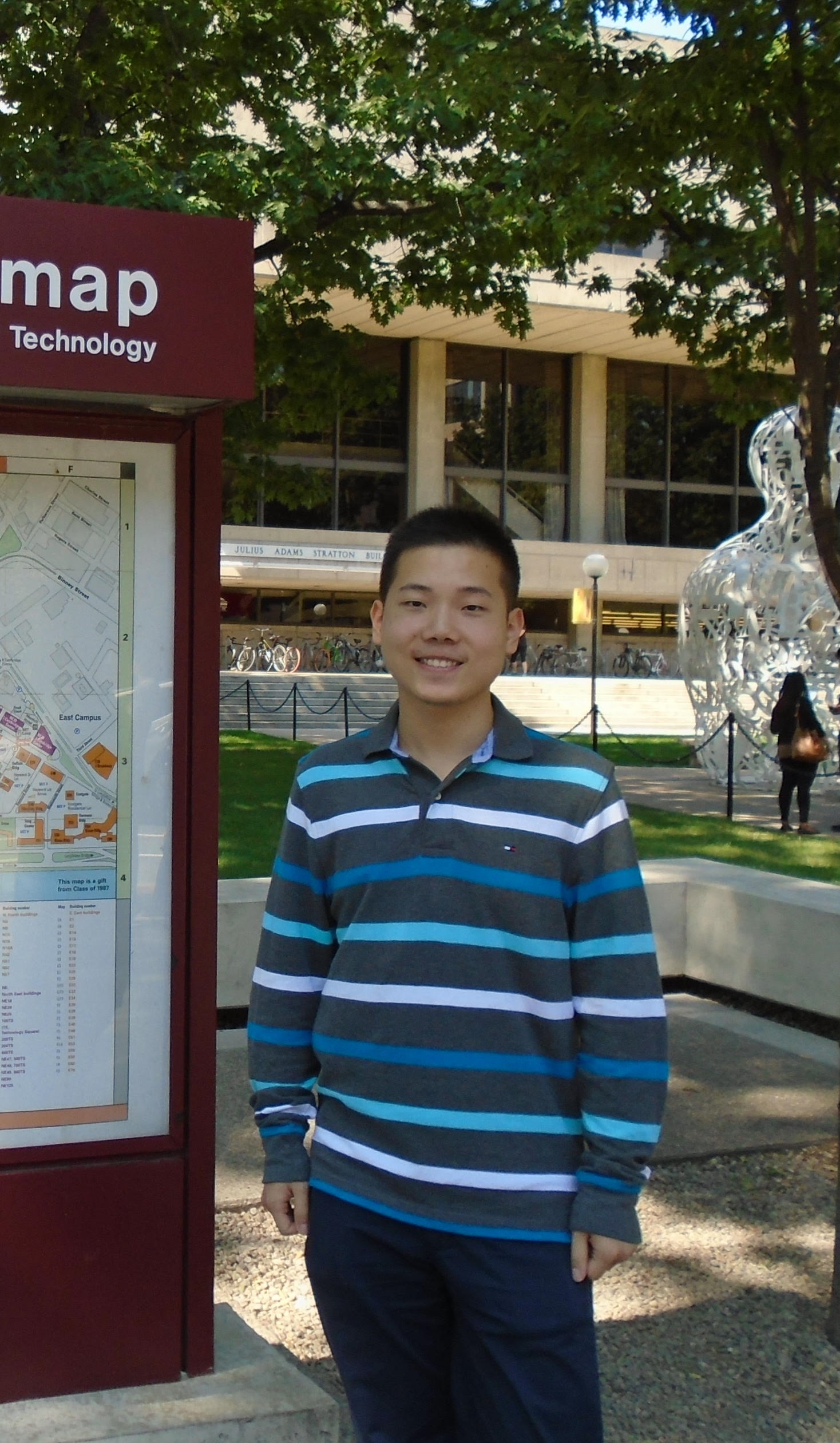
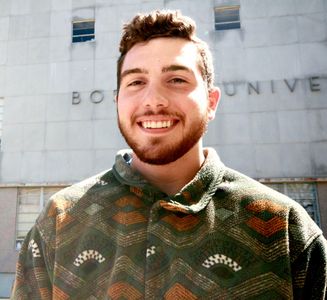
The 2018 Beckman Scholars are pictured below.

Andrea Cheng
Advisor: Dr. John Porco, Department of Chemistry
Synthesis of Asperterpene A and Related Meroterpenoids
The chemical synthesis of natural products and their derivatives in the laboratory has provided a means to create large quantities of drugs of biomedical importance. Meroterpenoids are natural products partially derived from terpene pathways. Numerous compounds derived from animals, plants, bacteria, and fungi belong to this class of natural products, which show a wide range of biological activities. For example, asperterpene A, a 3-5-dimethylorsellinic acid (DMOA)-based meroterpenoid, was isolated from the fungus Aspergillus terreus in limited amounts. Biological studies have shown that this compound is a potent β-secretase (BACE1) inhibitor. BACE1 is a protease enzyme that is responsible for the production amyloid β in the brain by cleavage of the amyloid precursor protein (APP). Therefore, BACE1 inhibitors are intensely being pursued in the treatment of Alzheimer’s disease (AD), which is caused by the accumulation of amyloid β in the brains of AD patients. This project, being carried out in the laboratory of John Porco (Chemistry Department, will primarily focus on the development of a synthetic pathway for the synthesis of asperterpene A and derivatives. The goals of the project include asymmetric synthesis of intermediates as well as cyclization studies to afford a variety of DMOA-derived meroterpenoids, including asperterpene A. Overall, it is hoped that this project will lead to the development of new classes of therapeutics for the treatment of Alzheimer’s disease.

Katie Tiemeyer
Advisor: Dr. Kim McCall, Department of Biology
Investigating the Impact of Protein Glycosylation on Glial Phagocytosis
Cell death and subsequent phagocytic clearance are essential processes for proper development and tissue homeostasis. In the brain, glial cells function as the primary phagocytes, responsible for the engulfment and degradation of apoptotic neuronal cell bodies. We have previously shown in Drosophila that the absence of Draper, a trans-membrane phagocytic receptor, results in an accumulation of neuronal cell corpses during larval development that persist in an unchanging quantity throughout adulthood. Furthermore, the absence of Draper ultimately yields agedependent neurodegeneration in adults. This phenotypic combination closely mimics human neurodegenerative disease progression. Therefore, we are interested in elucidating mechanisms that result in neural degeneration. To that end, increasing attention has been paid to understanding the role of cellular protein glycosylation profiles in regulating intracellular miscommunication and disruption of normal cell function. In order to more fully understand the role of protein glycosylation in the context of defective glial phagocytosis and neurodegeneration, we are exploring how lack of draper may alter glycoprotein modification processes. We have identified qualitative differences in nucleocytoplasmic O-linked N-acetyl glucosamine (O GlcNAc) protein modification and in cell surface HRP-epitope glycan modification in draper mutants. Our work is focused on identifying mechanistic links between altered glycoprotein glycosylation in neural
tissue and neurodegeneration mediated by defective glial phagocytosis.
2017 Beckman Scholars
Graduate May 2019

Megan Hoffman
Advisor: Dr. Adrian Whitty, Department of Chemistry
Characterization of Macrocyclic Compounds as Inhibitors of the Protein-Protein Interaction Between β-Catenin and TCF4
Protein-protein interactions (PPIs) play critical roles in essentially all intracellular signaling pathways, and therefore represent a significant portion of potential drug targets. Small molecules that follow classic drug design criteria have, however, proven ineffective at disrupting PPIs. Protein complexes often interact via relatively large, open binding surfaces, which lack binding pockets suitable to accommodate a typical druglike compound. We hypothesize that larger macrocyclic compounds (MC), defined as compounds with ring structures of at least 14 atoms, have the potential to inhibit challenging drug targets such as PPIs. Current success with natural product-derived MCs as drugs indicates therapeutic potential; however, purely synthetic MCs have not been widely investigated. It has been determined in preliminary work by the Whitty and Vajda Labs that the protein β-catenin, with its binding partner TCF4, is a suitable PPI target for MC inhibition. Computational analysis indicates that the sites on β-catenin that contribute the bulk of its binding energy with TCF4 are separated by too great a distance to be bridged by a classical drug-like compound; however, a large MC of appropriate design could span this distance. This project will primarily serve as a test case for the development of synthetic MCs as inhibitors of challenging PPI targets. Further characterization of the interactions energetics at the interface between β-catenin and TCF4 will confirm the strengths and locations of binding energy hot spots at the interface, guiding the design of MCs to exploit those sites. β-catenin, like other challenging PPI targets, is known to play a role in the regulation of cancer cell growth and proliferation. Therefore, the ability to disrupt the interaction between proteins using synthetic MCs could be enabling for future drug development.

Christopher Petty
Advisor: Dr. Frank Naya, Department of Biology
Investigating the Role of the Gtl2-Dio3 noncoding RNA locus in Cardiac Muscle Cell Differentiation
Heart disease is associated with widespread dysfunction and significant loss of cardiac muscle cells. Regardless of etiology, gene expression is radically altered suggesting that the cardiac muscle genome is reprogrammed in the disease process. Thus, characterizing the genomic mechanisms that govern cardiac muscle cell homeostasis, differentiation, and function is essential to our understanding of the pathological processes in heart disease. Recently, we reported that a noncoding RNA locus, named Gtl2-Dio3, functions in skeletal muscle differentiation and regeneration, and in cardiac muscle cell proliferation. The Gtl2-Dio3 noncoding RNA locus encodes a long noncoding RNA and a mega-cluster of more than 50 micro RNAs. Expression of the noncoding RNAs in the Gtl2-Dio3 locus was found to be coordinately dysregulated in mouse models of heart disease. Given these exciting results, we propose to delve deeper into the regulatory role of Gtl2-Dio3 noncoding RNA locus in cardiac muscle cells. This project will test the hypothesis that the Gtl2-Dio3 locus has an essential regulatory and epigenetic function in cardiac muscle biology and disease. Specifically, we will use CRISPR gene editing to mutate the Gtl2-Dio3 locus in mouse embryonic stem cells followed by differentiation to cardiac muscle cells in order to define the collective function of its noncoding RNAs in this process. The molecular dissection of noncoding RNAs in cardiac muscle cell differentiation will be an important step in the development of genomic strategies to treat heart disease.
2016 Beckman Scholars
Graduate May 2018

Grace Ferri
Advisor: Dr. Karen Allen, Department of Chemistry
Designing a Trehalose-6-Phosphate Phosphatase Inhibitor
Lymphatic filariasis is a mosquito-borne disease caused by infection from nematodes, or roundworms. Ninety- and ten-percent of lymphatic filariasis infections are caused by the parasites Wuchereria bancrofti and Brugia malayi, respectively. Although the pathogen tends to invade the host during childhood, disease, such as elephantiasis and river blindness, manifests during adulthood. At present, 120 million people are infected with lymphatic filariasis in the developing world. However, the currently approved treatment regimen of albendazole in combination with diethylcarbamazine citrate or ivermectin does not eliminate parasite infection until 4-6 years after administration.
The Allen laboratory has focused on the protein product of the gob1 gene as a potential target for antifilarial therapeutics. When RNA interference is directed at the gob1 gene in Caenorhabditis elegans (a widely used non-pathogenic model nematode), the knockdown of gene function results in 100% of the roundworms experiencing lethality attributed to larval arrest and gut obstruction within 24 hours. Prior research indicates the encoding of the enzyme trehalose-6-phosphate phosphatase (T6PP) by the gob1 gene. T6PP is a member of the haloalkanoic acid dehalogenase superfamily (HADSF) that acts upon trehalose 6-phosphate, which is the intermediate in the pathway by which glucose 6-phosphate is converted to trehalose for energy production and metabolism. In the absence of the T6PP gene product, trehalose-6-phosphate accumulates to a level toxic to the nematode.
The T6PP gene sequence is conserved not only within species of the genus Caenorhabditis but also among the porcine and human nematode pathogens including B. malayi. In addition, bacterial pathogens such as Mycobacterium tuberculosis utilize trehalose to synthesize cord factor, a mycolic acid-derived membrane glycolipid enabling toxicity, pathogenicity, and phagocyte survival. In M. tuberculosis, knockout of the otsB gene encoding T6PP precludes pathogenic growth in laboratory culture and virulence in a mouse model. Thus, the trehalose pathway affords an additional opportunity for intervention in bacterial pathogenesis.
Our laboratory has analyzed the X-ray crystallographic structures of numerous HADSF with respect to the conserved active-site platform. These studies reveal that a common ligand-induced closure of the “cap” domain upon the catalytic “core” domain lends substrate specificity. The structure of B. malayi T6PP has been obtained in our laboratory but a structure liganded to substrate or substrate analogs has not yet been achieved, nor have the structures of additional nematode and bacterial targets been determined. We aim to determine the X-ray crystal structure of M. tuberculosis T6PP and structures of B. malayi T6PP in the presence of substrate and/or substrate analogs. By comparing steady-state kinetics among species in which T6PP is conserved, performing structure-guided mutagenesis, and utilizing hot spot mapping, we seek to design a high-affinity T6PP inhibitor. Employing the HADSF drug target would provide remission from lymphatic filariasis on a much more rapid timescale than the alternative therapeutics on the market as well as deliver a new route to treatment of tuberculosis.

Jonathan Messerschmidt
Advisor: Dr. Thomas Gilmore, Department of Biology
The Evolutionary Origins of Toll-like Receptor Signaling
The innate immune system is the most primitive branch of animals defense against pathogens, and is important for eliciting responses such as inflammation and antimicrobial peptide production. Toll-like receptors (TLRs) are membrane-bound receptors that recognize microbial pathogens in a range of organisms from insects to humans. The discovery of TLRs in Drosophila and mammals was the basis for the Nobel Prize in 2011. Genome sequencing efforts have shown that TLRs exist in a number of organisms more basal to flies, but the biological roles of these TLRs for innate immunity or other processes is not known. Our lab has been characterizing a TLR-like molecule in the sea anemone Nematostella vectensis (Nv), which is a model system of the phylum Cnidaria that comprises a variety of mostly marine invertebrates, including anemones, corals, hydra and jellyfish. In preliminary results for this project, we have cloned and began to characterize the single TLR cDNA from Nematostella. The Nv-TLR encodes a transmembrane protein that has substantial sequence homology to human TLR4. Moreover, sequences from Nv-TLR can induce activation of transcription factor NF-kB in human cells. Using an antibody to Nv-TLR, we have found that Nv-TLR is expressed in a phylum-specific cell type called cnidocytes, which are the same cells that express Nv-NF-kB. Thus, these results suggest that a TLR-to-NF-kB pathway is conserved in cnidarians. The goals of my project include the following: 1) to identify sequences in Nv-TLR that are important for activation of NF-kB; 2) to find microbial compounds that can activate Nv-TLR-to-NF-kB signaling in mammalian cells and intact anemones; and 3) to elucidate downstream targets for Nv-TLR-to-NF-kB signaling in Nematostella. Overall, the outcomes of this research may reveal important information about the evolutionary basis of vertebrate innate immunity, and provide information on how environmentally threatened marine organisms respond to certain types of pathogenic stress.
2015 Beckman Scholars
Graduate May 2017

Anastasia Nizhnik
Advisor: Dr. Cynthia Bradham, CAS Biology
Molecular Characterization of Genes Specifying Skeletal Patterning in the Sea Urchin Model
Developmental patterning is a complex process that regulates tissue specification and morphogenesis. The sea urchin larval skeleton provides a comparatively simple model in which to study patterning of developmental events. Skeletal patterning in the sea urchin embryo requires cell-cell interactions between the pattern-containing ectoderm and the skeletogenic primary mesenchyme cells (PMCs). The Bradham lab previously performed a high-throughput screen to identify ectodermal patterning genes. This work identified SVEP and LOX as ectodermally expressed gene candidates for specifying skeletal patterning. SVEP encodes an adhesion protein that had not previously been characterized in the sea urchin. LOX is a gene whose product, lipoxygenase, acts on arachidonic acid to produce HETES, which in turn impacts multiple signal transduction pathways including PI3K, PKC, and Ras signaling. A genetic loss-of-function (LOF) analysis for either SVEP or LOX led to dramatic defects in the orientation of the initial skeletal elements, which are known as triradiates. The purpose of this project is to understand how the genes SVEP and LOX work together to orient the larval skeleton. Our goal is to perform combined LOF for SVEP and LOX to determine if the triradiate orientation defects observed in the individual LOF studies are additive or synergistic. That is, the scholar will investigate skeletal morphology, PMC positioning and triradiate orientation in double LOF embryos (i.e., embryos with SVEP and LOX simultaneously inactivated). The results of this project will establish the function of SVEP and LOX in sea urchin embryonic skeletal patterning. Understanding the molecular mechanisms underlying skeletal pattern formation in sea urchin embryos will likely uncover conserved patterning genes and their functions. Interestingly, many of the skeletal patterning candidate genes identified in the Bradham lab have been generally implicated in developmental patterning as well as cancer migration and metastasis. Thus, a more complete understanding about how these sea urchin genes function to direct cell migration and skeletal patterning will provide information about developmental processes, and will likely also provide insight into cancer progression.

Nicole Carter
Advisor: Dr. Tom Gilmore, CAS Biology
Invertebrate Models to Understand the Molecular Basis of Immunity and Symbiotic Relationships
Almost all organisms, including humans, have symbiotic relationships with other organisms, which are essential for the health of both organisms. To cite one prominent example, the health of coral reefs often relies on symbiotic relationships between corals (members of the phylum Cnidaria) and photosynthetic algae, and these symbiotic relationships can be interfered with by external stresses (ocean acidification, warming, etc.). However, little is known at the molecular level about how these relationships are maintained, or disrupted. This project, which is being conducted in collaboration with the lab of Virginia Weis at Oregon State University, seeks to deepen our understanding of the molecular processes that occur during symbiosis in simple marine ecosystems. Specifically, we are using a model cnidarian, the anemone Aiptasia pallida to explore symbiotic relationships involving the anemone and a series of symbiotic algae. Specifically, the project aims to characterize activation of the NF-κB transcription factor pathway in Aiptasia in response to symbiont introduction or escape. NF-κB is well-known for its role in immunity from flies to humans, and based on preliminary results from Aiptasia, it is a reasonable hypothesis that NF-κB is also be involved in host-symbiont or host-pathogen responses in lower organisms. The Gilmore lab has shown that cnidarians including Aiptasia contain NF-κB, but its exact role in marine cnidarians is only poorly understood, NF-κB. For this project, I have cloned the cDNA encodingAiptasia, and have conducted preliminary experiments towards characterizing the protein, Ap-NF-κB. Using cell- and lab-based model systems, I will characterize effects of environmental stress and the consequent loss of symbiosis on NF-κB activity. Overall, the outcome of this research will likely reveal the evolutionary basis of vertebrate immunity, and provide information on the effects of environmental stress on molecular pathways in sensitive marine organisms.
2014 Beckman Scholars
Graduated May 2016

Shannon Anderson
Graduated May 2016
Advisor: Dr. Joyce Wong, ENG Biomedical Engineering
Biomaterial Scaffolds for Nerve Repair
One of the most rapidly expanding areas of biomedical research is regenerative medicine and biomaterials. Biomaterials can be both naturally derived and artificially created, and have a variety of uses, one of them being the scaffolding of bioengineered tissues. Scaffolding biomaterials, which provide the framework for regenerating tissue, will be a focus of this project. The natural polymer, silk, will be looked at in particular depth. Silk has a variety of applications in biomedical research due to its highly desirable properties such as its high tensile strength as well as its biocompatible and biodegradable properties. The silk will be created using microfluidics technology. This highly controlled process most closely mimics natural silk spinning and appropriately replicates the key structural attributes of native silk. The silk will be analyzed using both experimental and computational approaches; a combination which allows for a higher control over the properties and functionality of the biomaterial produced. Among the vast applications of silk, the area this project will focus on is its use in scaffolding for nerve repair and regeneration. Previously, nerve repair has been studied using autograft techniques, which utilizes other tissues taken from the patient’s own body as a scaffold. However, it has been seen that there are considerable benefits to using artificial nerve guide conduits (NGCs) for the purpose of nerve regeneration after an injury. Finding an appropriate biomaterial for artificial NGCs is difficult due to the need for them to be flexible, yet strong enough to resist compression, as well as ideally biodegradable. Silk’s strength and biological properties make it a promising choice as an artificial NGC. In the long-term, this project can have a lasting impact on the treatment of central nervous system injuries, including the potential to restore motor function by allowing for the reestablishment of lost neural connections. Shannon is current in a PhD program in Biomedical Engineering that is jointly run by the Georgia Institute of Technology and Emory University.

Mikayla Huestis
Graduated May 2016
Advisor: Dr. Adrian Whitty, CAS Chemistry
Quantitative Investigation of RET Activation and Signaling
Despite decades of research on the activation and signaling of growth factor (GF) receptors, our current understanding of these processes remains largely qualitative. Our goal is to advance our quantitative, mechanistic understanding of how GF receptors mediate the ability of cells to sense and respond to their environment with RET as our investigational system. RET is a receptor tyrosine kinase that mediates the response of certain neuronal cells to a family of growth factors comprising GDNF, neurturin, artemin and persephin. Previously we have used conventional biochemical and cell biological assay methods to characterize RET activation and signaling. However, to advance our research program we need access to more sophisticated methods, especially those that enable single cell measurements by microscopy and/or flow cytometry. An initial goal will be to create two different types of engineered cell lines, to serve as quantitative reporters of early and late events in RET signaling, and to use them to address questions concerning how activation of RET by artemin is quantitatively coupled to gene transcription. One goal will be to develop a cell line in which RET is fused to mutant forms of green fluorescent protein, known as mCer and mYPet, designed to give an optical signal when two RET molecules interact on the cell membrane, using the method of Förster Resonance Energy Transfer (FRET). A second goal will be to generate a luciferase reporter gene cell line to provide a convenient and quantitative read-out of RET-dependent activation of gene transcription. Assuming at least one of these cell line engineering efforts is successful, we will then move on to use the newly developed tool to address fundamental questions concerning quantitative aspects of RET activation and signaling. Mikayla is current in an MD program at the Boston University School of Medicine.
2013 Beckman Scholars
Graduated May 2015

Audrey Lambert
Advisor: Dr. Tom Gilmore, CAS Biology
Genetic Modification of the Starlet Sea Anemone, Nematostella vectensis
A key scientific advance for understanding basic biological processes has been the ability to genetically alter model organisms, such as nematodes, fruit flies, and mice. The phylum Cnidaria includes approximately 10,000, mostly marine, basal organisms, including several that are of commercial importance or are under environmental insult, esp. corals. An emerging model organism for animals in this phylum (corals, anemones, jellyfish, hydra) is the starlet sea anemone Nematostella vectensis. This anemone is convenient because it is small, can be collected from natural estuarine sites, is easily maintained in the lab, has extensive regenerative properties, and has a complete genome sequence and extensive RNA expression data. Nevertheless, molecular genetic approaches to modify gene function are limited in this animal. Thus, a major goal of this project is to develop additional and improved methods for altering and introducing gene function in Nematostella. These techniques will then be used to study signaling pathways (e.g., the NF-κB pathway) and biological functions, and perhaps to develop biosensor or decorative animals. For example, one long-term goal may include developing transgenic N. vectensis as biological sensors for environmental factors such as pH, temperature, and salinity. Audrey is currently in a combined MSW/MPH program at the Boston University School of Medicine.

Sarah Nocco
Advisor: Dr. Frank Naya, CAS Biology
Mapping Transcriptional Enhancers of Mef2a in Skeletal Muscle Differentiation and Regeneration
There are currently no effective treatments for the various forms of muscular dystrophy. The main form of ‘treatment’ consists mainly of aiding patients in controlling the symptoms that accompany their disease. A promising approach in the treatment of muscular disorders involves repairing or replacing diseased muscle with healthy muscle by means of regeneration. Skeletal muscle is one of the few mammalian tissues that has the capacity to regenerate, with new cells arising from muscle stem cells residing in proximity to mature muscle fibers. While many molecular pathways have been identified that regulate muscle regeneration, our understanding of this process is far from complete. Recently, it has been found that transcription factor MEF2A is a key player in the regulation of skeletal muscle regeneration after injury. Given the importance of MEF2A in muscle regenerations, it is essential to understand the mechanisms by which MEF2A is itself regulated because such information will provide insight into the molecular control of muscle regeneration. Specifically, we propose to investigate the transcriptional regulation of the Mef2a gene since its expression is modulated in skeletal muscle regeneration. Our goal is to identify transcriptional enhancers of Mef2a by performing deletion mutagenesis of the proximal promoter and analyzing the activity of these sequences using reporter assays. Various promoter deletions will be analyzed both in vitro in an established muscle cell line and in vivo using a muscle regeneration model in mice. The minimal cis-acting DNA sequences will then be used to identify upstream transcriptional regulators and signaling pathways that control the expression of Mef2a. Overall, these studies will enable us to develop a more complete understanding of the pathways employed in skeletal muscle regeneration, which may have therapeutic relevance for human muscle degenerative diseases. Sarah is currently in an MD program at the Boston University School of Medicine.
2012 Beckman Scholars
Graduated May 2014

Alex Valentine, a Biomedical Engineering major, conducted research with Dr. Joyce Wong (ENG Biomedical Engineering) on the development of a drug delivery technique for the treatment of allergic asthma and atopy. The project seeks to develop an ultrasound-facilitated transdermal antibody delivery method that can be validated in mouse models. This project has potential medical implications in that the specific antibody being used has shown therapeutic benefits for over half of asthma patients who have a particular allergen imbalance.

Zach Ariki, a Chemistry major, conducted research with Dr. John Porco (CAS Chemistry). The general area of his research is the organic synthesis of natural product-based compounds that may have medical benefits. Specifically, Zach is investigating the synthesis of a class of compounds called microsphaerins, which have been demonstrated to have antimicrobial activity against methicillin-resistant Staphylococcus aureus (MRSA), which is now a common hospital pathogen for which there are limited therapeutic options. Thus, the development of methods to synthesize microsphaerins could have important benefits for the treatment of patients with MRSA. Zach is currently a PhD student in Chemistry at Queens University in Ontario, Canada.
2011 Beckman Scholars
Graduated May 2013

Claire Schenkel, a Biology (specialization in Cell Biology, Molecular Biology & Genetics) and Speech, Language & Hearing Sciences double major, performed research with Dr. Kim McCall (CAS Biology). Claire’s research focused on the role of programmed cell death in the ovary using the model organism the fruit fly Drosophila melanogaster. This work has relevance to understanding reproductive physiology, development and certain disease processes. Claire is currently in a joint Harvard-MIT PhD program in Speech & Hearing Sciences.

Rishikesh Kulkarni, a Biochemistry & Molecular Biology major, and performed research with Dr. Sean Elliott (CAS Chemistry). He conducted biochemical research to understand the role of iron-sulfur clusters in a novel class of proteins called CDGSH proteins. Using computational protein similarity network analysis, Rishi sought to understand the functional and evolutionary relationship between this new class of proteins, some of which appear to play a role in human diabetes. Richi is currently in a PhD program in Chemistry at the University of California, Berkeley.
2010 Beckman Scholars
Graduated May 2012

Colleen Drapek, a Biochemistry & Molecular Biology major, conducted research with Dr. Frank Naya (CAS Biology) on the characterization of the regulatory mechanism of the transcription factor MEF2A by the gene BMP2. This work will have relevance to the understanding of cardiovascular development and disease. Colleen Drapek is currently in a PhD program in Developmental & Stem Cell Biology at Duke University.

Angela Xie, a Biomedical Engineering major, performed research with Dr. Joyce Wong (ENG Biomedical Engineering) on characterizing the mechanical properties of mesenchymal stem cell sheets for tissue engineered vascular patches. This work will have applicability to the development of artificial tissues, such as blood vessels. Angie is currently in a PhD program in Biomedical Engineering at the University of Wisconsin.
2009 Beckman Scholars
Graduated May 2011

Meredith Duffy, a Biomedical Engineering major, performed research with Dr. Joyce Wong (ENG Biomedical Engineering) and used tissue engineering approaches to develop a small-diameter artery replacement by combining cell sheet engineering with microtexturing techniques.

Arthur Su, a Biochemistry & Molecular Biology major, performed research with Dr. John Porco (CAS Chemistry) on the organic synthesis of natural product derivatives that may have anti-bacterial and anti-cancer cell growth properties.
2008 Beckman Scholars
Graduated May 2010

Florencia Rago, a Biochemistry & Molecular Biology major, performed research with Dr. Dean Tolan (CAS Biology) on enzyme protein structure and function. Florencia Rago completed her PhD in Biology at MIT, and is currently a post-doctoral fellow at Novartis.

Tyler Ford, who was a Biochemistry & Molecular Biology major, worked with Dr. Tom Gilmore (CAS Biology) performing research on the molecular basis of lymphoma. Tyler completed his PhD at Harvard and is currently a scientist at Addgene in Cambridge, MA.
The previous Beckman Program Award supported six students.
The 2006 Beckman Scholars performed research in CAS Biology with Dr. Tom Gilmore (Melissa Chin), Dr. Dean Tolan (Michael Coyle) and Dr. William Eldred (James Flannery). These scholars graduated in May 2008.
The 2005 Beckman Scholars were Beth Cimini, Daniel Bruggemeyer, Roy Arjoon. These scholars graduated in May 2007.