Building Brain Complexity
Unlocking neurons’ secrets at the earliest stage of development
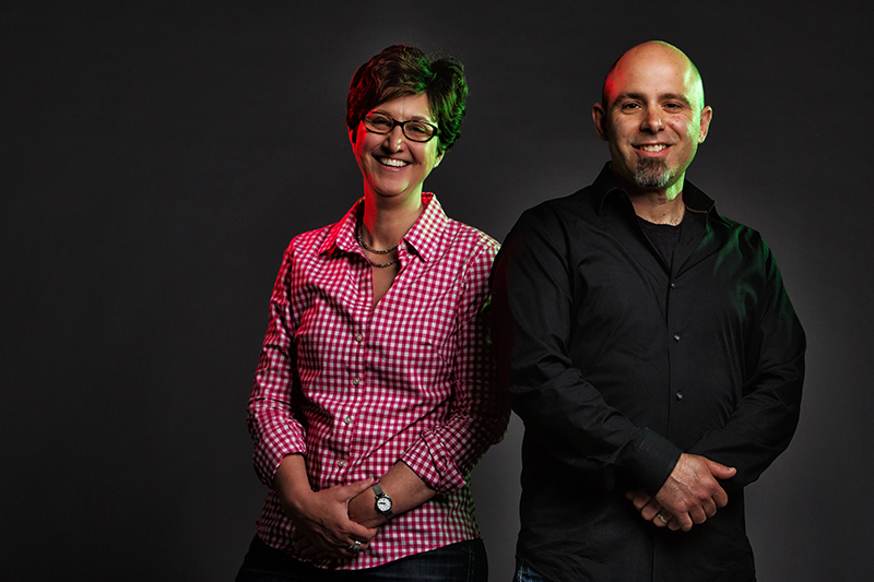
Jennifer Luebke and Tarik Haydar, associate professors of anatomy and neurobiology, study how stem cells develop into neurons. “If we have a better understanding of how the brain works under normal conditions, then we can have a better understanding of how it works when things go wrong,” says Luebke. Photo by Michael D. Spencer
The cerebral cortex is what most people imagine when they think of the word “brain.” It’s the convoluted mass of gray matter that’s responsible for memory, thought, language, consciousness—things that make us unique. “It’s the seat of all our higher functions,” says Tarik Haydar, a Boston University School of Medicine (MED) associate professor of anatomy and neurobiology. “Understanding how it comes together is part of the challenge and the excitement of our work.”
Haydar and his colleague Jennifer Luebke (GRS’90), MED associate professor of anatomy and neurobiology, are trying to understand how and when the complexity of the cerebral cortex is first established. Their discoveries, published in the Journal of Neuroscience in April 2015, show that this process begins when stem cells generate neurons even before we are born. Their work offers the first direct evidence that distinct lines of neurons are generated from different types of neural stem and progenitor cells, and that these diverse neurons populate regions of the brain once thought to be homogenous. The work, funded by the National Institutes of Health (NIH) and the Rafael del Pino Foundation, has widespread implications for our basic understanding of the brain, especially against the backdrop of the European Union’s Human Brain Project (HBP) and the United States’ BRAIN Initiative, the goal of which is to map our neural circuits in fine detail.
“If we have a better understanding of how the brain works under normal conditions, at the most basic level, then we can have a better understanding of how it works when things go wrong,” says Luebke. “Why are some neurons more vulnerable to Alzheimer’s and Parkinson’s diseases? We need an understanding of how neural diversity is generated to answer these questions.”
The human brain contains billions of neurons, all slightly different from one another. Why they are different and how they got that way are some of the current puzzles of neuroscience. Brain growth starts early in fetal development. At this stage, neural stem cells—think of them as the grandparents of your brain cells—differentiate into a handful of intermediate “progenitor” cells, akin to parents. The various progenitors then give rise to the end products: neurons. And the neurons, like human children, are somehow all different. But when do these differences first arise? And how? “Our molecular understanding of the neural progenitors has changed dramatically in the past 10 years or so. Using this new knowledge, we were able to ask focused questions about how they work,” says Haydar. “We now know there isn’t just one type of progenitor cell in the brain, and particularly in the cerebral cortex, but there are half a dozen, maybe even more. We wondered why there are so many types of progenitors. What’s their purpose and function?”
For many years, scientists theorized that we had so many different progenitors because more progenitor cells meant more neurons, leading to our big human brains. “That sort of made sense,” says Haydar, “but it turns out that all the progenitor types in the really complicated human brain are also found in the simpler mouse brain. So it seemed that something else might be going on.”
Haydar’s group wondered if the many types of progenitor cells led not to more neurons, but to different kinds of neurons. To test the idea, they needed a way to build a genealogy of the brain, tracing which progenitor cells led to which neurons. Then they could test the descendants for differences. Bill Tyler, a postdoctoral fellow in Haydar’s lab, created the tools to do it.

Tyler focused on one type of cell called the basal intermediate progenitor, which was thought for many years to give rise to most of the neurons in the upper layers of the cortex. These particular cells express a gene called T-box brain protein 2 (TBR2) while other progenitors in the fetal brain don’t. Tyler used this to his advantage, designing a genetic tagging technique that labeled basal intermediate progenitors in fetal mice with a fluorescent protein called mCherry. That way, the progenitors and all of their descendant cells would glow red. At the same time, Tyler’s method marked all the other progenitor cells green with a fluorescent protein called ZsGreen. Then he waited until the mice were born, and looked at their brains with a laser microscope, which gave him the high resolution needed to see tiny details of the neurons.
The picture he saw revealed that the astounding complexity of the cortex was now slightly ordered by the two fluorescent proteins: red and green brain cells popped brightly against a black background, their colored tendrils snaking off in all directions. But besides the obvious color difference, were the red and green neurons different in any other way? If so, how? “So what we did then was go into the brain and measure the precise shapes and activities of the red and green neurons,” says Haydar.
That’s where Luebke entered the picture. Her lab specializes in neuron electrophysiology—measuring the electrical output of single brain cells. It’s a challenging process. “It’s not that easy to get the readings. A lot of things have to go right,” says Luebke. “Each recording takes about an hour. Sometimes you think you’re on a cell, and then you’re not. In any given animal, we’d only be able to record from three or four cells that could be subsequently reconstructed.”
The painstaking teamwork paid off with some remarkable results—they discovered that the red and green neurons were strikingly different. “The red cells were much more excitable,” says Luebke. Although the red neurons were physically simpler than their green cousins, they fired off many more electrical impulses when stimulated. The cells also differed in their “input resistance,” which strongly influences their response to an incoming electrical signal. “The red neurons and the green neurons were very different from one another, even though they were all born at the same time and were going to the same place,” says Haydar. “I think what this shows is that there are fundamental components of a neuron’s identity that are formed at very early stages of development.”
The scientists think that this ability of stem cells to generate early differences between neurons plays a large role in the how the healthy brain functions. One of their next steps is to see whether changes in progenitors may cause cognitive problems in specific human disorders. Tyler and Haydar recently showed that one type of progenitor cell is reduced in the fetal brain of a mouse model of Down syndrome. These changes could mean that neuron diversity may be altered at very early stages of life, and that this disorder may take root in the brain before neurons are even made. A deeper understanding of this basic science may one day lead to better treatments for developmental disabilities.
“The brain is a vast number of interconnected circuits, and we’re just at the very beginning of understanding how the circuits work, let alone how they come to be,” says Haydar. “But this work opens up a new understanding and a brand new arena of research.”

Comments & Discussion
Boston University moderates comments to facilitate an informed, substantive, civil conversation. Abusive, profane, self-promotional, misleading, incoherent or off-topic comments will be rejected. Moderators are staffed during regular business hours (EST) and can only accept comments written in English. Statistics or facts must include a citation or a link to the citation.